
|
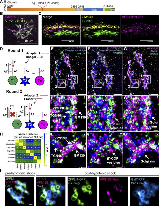
Fig. 1 VPS13B is localized at the interface between cis-Golgi and trans-Golgi membranes. (A) Domain cartoon of human VPS13B. (B) COS7 cells expressing codon-optimized human VPS13B^GFP immunolabeled for GFP and GM130. Scale bar = 10 µm. (C) Cross-section (side view) of HeLa cells expressing codon-optimized human VPS13B^GFP immunolabeled for GFP, GM130, and TGN46 imaged using 4Pi SMS microscopy. Scale bar = 500 nm. (D) Schematic representation of labeling steps used for FLASH-PAINT. FLASH-PAINT performed in HeLa cells expressing VPS13B^GFP and immunolabeled with anti-GFP. (E–G) FLASH-PAINT signals of a Golgi complex immunolabeled for the indicated Golgi complex targets. Scale bar = 2 µm. High magnification fields of the boxed areas in fields E–G are shown below in fields E′–G′ and E″–G″. Scale bar = 250 nm. (H) Median distances between super-resolved signals of different targets. Only signals closer than 500 nm to each other were considered. (I) Snapshots of COS7 cell expressing VPS13B^halo, ZFPL1-GFP (a cis-Golgi marker), and GalT-RFP (a trans-Golgi marker) before and after (8 min) hypotonic shock. Scale bar = 1 µm.