
Image
|
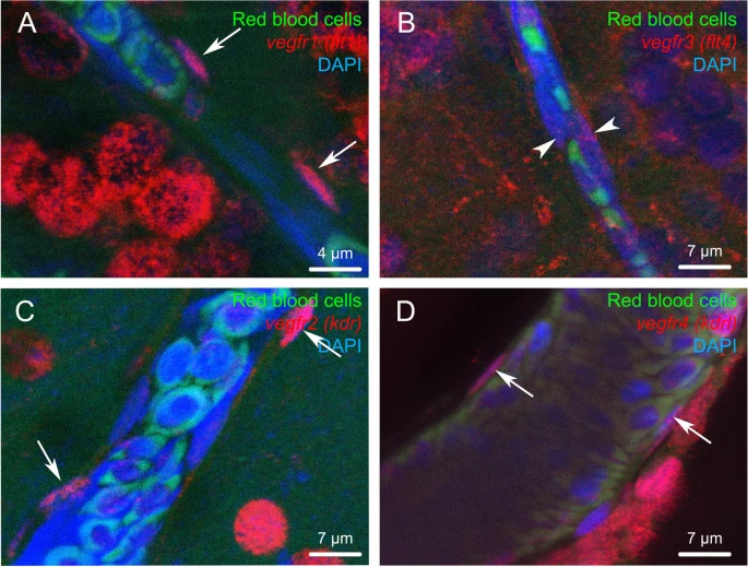
Figure Caption
Fig. 9 Vegf receptor gene expression in endothelial cells. Fluorescent in situ hybridization of vegfr (red) showing expression in endothelial cells for vegfr1, 2 and 4. Note that endothelial cells correspond to cells with an elongated nucleus (arrows) localized along the blood vessels. Arrowheads show a very weak vegfr3 labeling in endothelial cells. The red blood cells exhibit green autofluorescence. DAPI counterstaining allows to visualize cell nuclei (blue). Bars: 4 μm (A), 7 μm (B, C and D)
Acknowledgments
This image is the copyrighted work of the attributed author or publisher, and
ZFIN has permission only to display this image to its users.
Additional permissions should be obtained from the applicable author or publisher of the image.
Full text @ Neural Dev.