
|
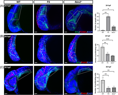
Fig. 8 fbln2 knockout causes increased apoptosis and decreased proliferation of CNCCs. (A) Terminal deoxynucleotidyl transferase dUTP nick end labeling (TUNEL) assay of embryos at 24 h post fertilization (hpf) revealed increased apoptotic cells in the dorsal cranial region of F0 and fbln2−/− (magnification factor: 20×, n = 30 per group, bar = 50 μm). The white dashed area highlights the distribution of CNCCs at the pharyngeal arches. Three wild type, three F0, and four fbln2−/− embryos were used for statistics. (B and C) Phosphorylated histone H3 (PHH3) staining revealed decreased proliferation in the craniofacial region and pharyngeal arches of mutant embryos, respectively, at 24 and 30 hpf (magnification factor: 20×, n = 30 per group, bar = 50 μm). The white dashed area highlights the pharyngeal arches. For statistics, six wild type, three F0, and six fbln2−/− embryos at 24 hpf, and three wild type, five F0, and six fbln2−/− embryos at 30 hpf were used. *p < 0.05; ***p < 0.001; ****p < 0.0001. Abbreviation: WT, wild type.