
|
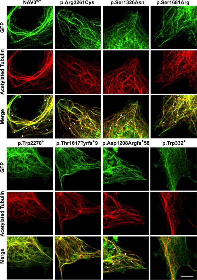
Fig. 3 NAV3 interacts with microtubules in COS7 cells. GFP-tagged WT and mutated NAV3 proteins (green) showed an overlap expression (yellow) with microtubules, decorated with acetylated tubulin antibodies (red). Granular expression of NAV3 was observed at dendrite-like structure formations and tip ends of microtubules. White arrow heads show the granule type expression at +Tip ends of microtubules. Similar to WT protein, the p.Arg2261Cys, p.Ser1326Asn, and p.Ser1681Arg variants harboring NAV3 also showed overlapping expression with stable microtubule structures. However, the protein-truncating variants, except p. Asp1208Argfs*58, did not show +Tip granule formations. Among the variants analyzed, NAV3 with p.Thr1617Tyrfs*9 and p.Trp332* had reduced or no bundling and show centralized expression in nucleus. Scale bar: 20 um.