
|
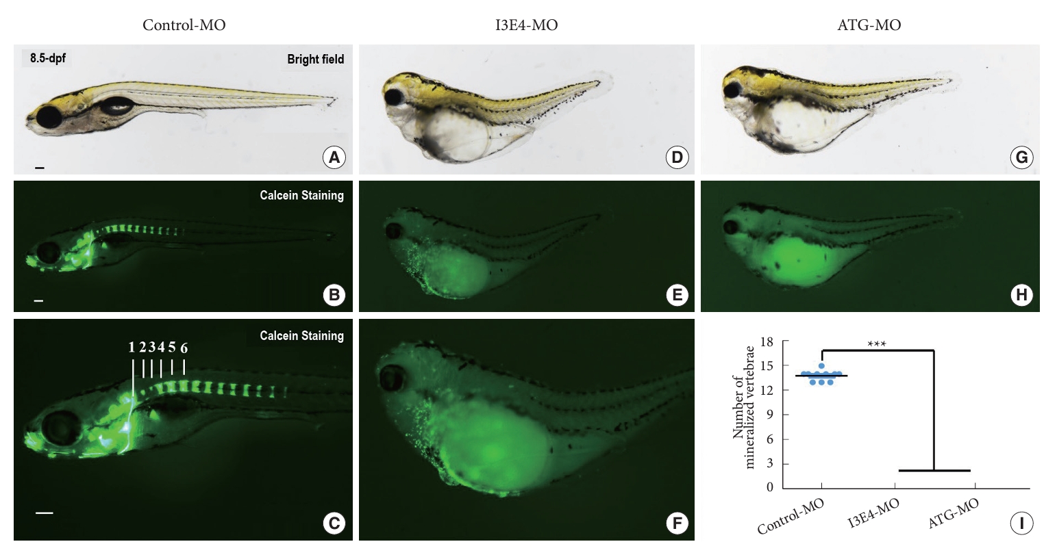
Fig. 4 Loss of best1 causes abnormal craniofacial structure and vertebral development. (A–H) Representative bright field and fluorescent images of zebrafish skeleton at 8.5-dpf. In vivo visualization of the skeleton is achieved by the administration of a fluorescent dye (Calcein) directly to the fish water. Dyes that bind to calcified matrix can be used to label the entire skeleton. Lateral view (B, C) of the skeleton and mineralized vertebrae of day-8.5 embryos labeled with Calcein. Vertebrae 1, 2, 3, 4, 5, and 6 are indicated. (E, F, and H) When embryos injected with best1-morpholino (MO) at one-cell stage, the amount of stained mineralized tissue is markedly reduced compared to control-MO-injected fish. Vertebral development is significantly delayed in best1 morphants. (I) Quantification of the number of mineralized vertebrae at 8.5-dpf. Columns, mean; standard error of the mean (n = 10; analysis of variance), ***p < 0.0001. Scale bar, 100 μm. dpf, days postfertilization.