IMAGE

Fig. 13.

ID
ZDB-IMAGE-240129-147
Source
Figures for Koosha et al., 2023
Image
Figure Caption

Fig. 13.

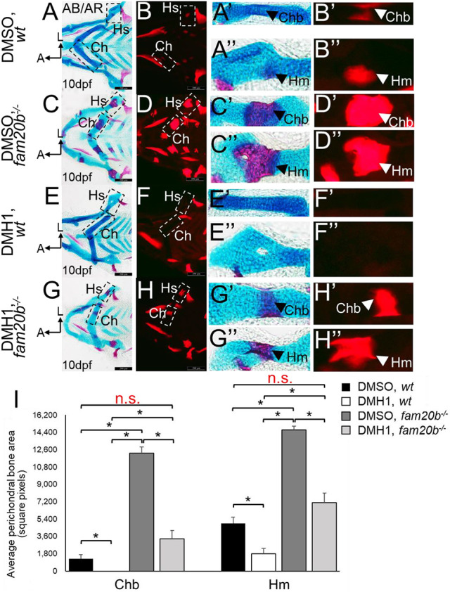
Inhibition of BMP signalling by early DMH1 treatment reduces perichondral bone formation in fam20b mutants down to wild-type levels at 10 dpf. (A-H″) Compared with DMSO-treated wild-type controls (A-B″), perichondral bone appeared to decrease in DMH1-treated wild types at 10 dpf (i.e. 6 days after end of 48 h treatment; E-F″). Similarly, perichondral bone at 10 dpf appeared to decrease in DMH1-treated fam20b mutants (G-H″), compared with DMSO-treated fam20b mutants (C-D″), with levels similar to that seen in DMSO-treated wild types (A-B″). (I) Quantitation of 20 embryos for each experimental group confirmed significant decreases (*P<0.05, one-way ANOVA and paired Student's t-test) in perichondral bone of DMH1-treated wild-type or fam20b−/− embryos. Remarkably, the levels of perichondral bone in DMH1-treated fam20b mutants was statistically indistinguishable from DMSO-treated wild-type controls. Scale bars: 200 μm (A-H). A, anterior; AB/AR, Alcian Blue/Alizarin Red; Ch, ceratohyal cartilage; Chb, ceratohyal bone; Hm, hyomandibular bone; Hs, hyosymplectic cartilage; HS, heat-shocked; L, lateral; n.s., not significant; wt, wild type.

Acknowledgments
This image is the copyrighted work of the attributed author or publisher, and ZFIN has permission only to display this image to its users. Additional permissions should be obtained from the applicable author or publisher of the image. Full text @ Development