IMAGE

Fig. 5.

ID
ZDB-IMAGE-240129-115
Source
Figures for Koosha et al., 2023
Image
Figure Caption

Fig. 5.

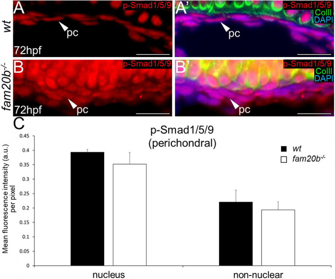
Smad-mediated BMP signalling does not increase in the fam20b−/− perichondrium region. (A-B′) p-Smad1/5/9 immunoreactivity did not show a clear difference in fam20b−/− perichondral cells (B,B′) at 72 hpf, compared with wild types (A,A′). (C) Quantitative image analyses (n=3 for each group) revealed no significant differences in p-Smad1/5/9 levels of fam20b−/− perichondral cells. Scale bars: 50 μm. a.u., arbitrary units; ColII, Collagen type II; pc, perichondral cells; wt, wild type.

Acknowledgments
This image is the copyrighted work of the attributed author or publisher, and ZFIN has permission only to display this image to its users. Additional permissions should be obtained from the applicable author or publisher of the image. Full text @ Development