
|
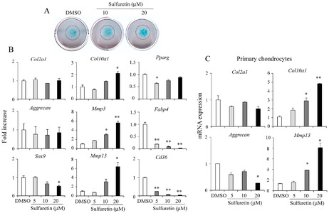
Fig. 1 Sulfuretin stimulates chondrocyte differentiation in cell culture. (A) C3H10T1/2 cells were differentiated into chondrocytes in the presence of sulfuretin (10 or 20 μM) for 12 days followed by Alcian Blue staining. (B) Relative mRNA expression levels of early chondrocyte genes (Col2a1, Aggrecan, and Sox9), late chondrocyte genes (Col10a1, Mmp3, and Mmp13), and adipocyte genes (Pparg, Fabp4, and Cd36) after sulfuretin treatment were determined by real-time PCR. (C) Primary chondrocytes were isolated from mice at postnatal day 7 and treated with sulfuretin for 12 days. Relative mRNA expression was quantified by real-time PCR. Data shown represent mean ± SEM. Statistical significance was determined relative to a control by Student’s t-test (*P < 0.05; **P < 0.01).