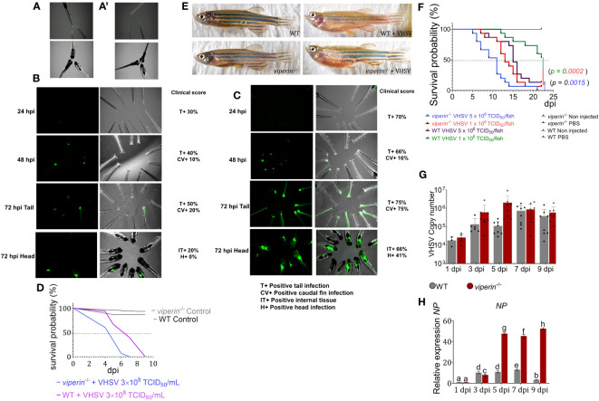
IMAGE

Figure 1

ID
ZDB-IMAGE-240105-44
Source
Figures for Shanaka et al., 2023
Image
Figure Caption

Figure 1

The VHSV infection experiment in viperin-/- zebrafish. rVHSV immersion experiment for larval zebrafish. (A) WT control larvae, (A’)viperin-/- control larvae. (B) WT, (C)viperin-/- fish were injured by caudal fin amputation method and then immersed in rVHSV. Migration of virus was analyzed after 24, 48, and 72 h postinfection (hpi). According to the experiment, rVHSV rapidly migrated into the internal tissue region of viperin-/- fish compared to WT fish. A clinical score represents the quantitative analysis of pathogenic symptoms. (D) Mortality recording for injury immersion experiment, according to the analysis, the viperin-/- fish had significantly higher mortality. (E) Morphology under VHSV infections. Adult WT and viperin-/- fish showed no differences in their morphology without VHSV infections. When infected with VHSV, both fish showed abnormalities such as edema, hemorrhage, and spinal defects causing head protrusions above the body axis (14 dpi). (F) A mortality experiment was conducted for the viperin-/- fish (n = 15/group). Two doses of VHSV were intraperitoneally injected into the fish (high dose 5 × 10⁶ TCID50/fish and the low dose 1 × 10⁶ TCID50/fish). Fish mortalities were recorded from 4 dpi onward. Mortality data were analyzed by the Kaplan–Meier (KM) method, and statistical significance was plotted using the Mantel–Cox test (p < 0.05). (G) VHSV copy number between the VHSV-injected viperin-/- and WT fish over time is shown, indicating a higher VHSV copy number in the viperin-/- fish. NP gene (H) expression in the viperin-/- fish shows significantly elevated levels compared to that in WT fish. VHSV, Viral hemorrhagic septicemia virus; rVHSV, Recombinant viral hemorrhagic septicemia virus; WT, wild type: dpi, days post infection; NP, VHSV Nucleoprotein.

Acknowledgments
This image is the copyrighted work of the attributed author or publisher, and ZFIN has permission only to display this image to its users. Additional permissions should be obtained from the applicable author or publisher of the image. Full text @ Front Immunol