
|
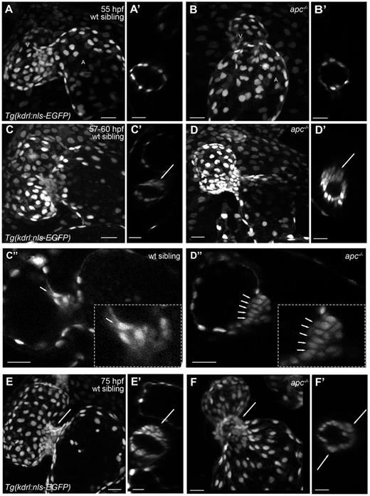
Fig. 6 apc mutants exhibit a disorganized accumulation of cells at the AVC. Maximum projection images of Tg(kdrl:nls-EGFP) apc mutant (B,D,F) and wild-type sibling (A,C,E) hearts at 55 (A,B), 60 (C,D) and 75 (E,F) hpf. Compared with the wild-type sibling hearts, the apc mutant hearts appear elongated (A-D). (C″,D″) Sagittal planes of 60 hpf wild-type sibling and apc mutant hearts, respectively. Insets in C″ and D″ show a magnification of the immature valve. At around 60 hpf, the valvular structures appear enlarged in apc mutants and more cells exhibit a cone-shaped nucleus (C″,D″, arrows). Note that this valve phenotype appears to depend on the overall phenotype of the heart; as the heart deteriorates the valvular structures at the AVC seem to dissolve. At 75 hpf, the distinctly structured immature valve leaflet in wild-type siblings (E,E′) is replaced by a disorganized accumulation of cells at the AVC of apc mutants (F,F′). Transverse planes through the AVC of apc mutants (A′-F′) show that these animals exhibit a more circular shaped AVC (B′,D′,F′), instead of the elliptically shaped AVCs seen in wild-type siblings (A′,C′,E′). Cells at the abluminal side of the valve appear to be distributed all around the AVC in apc mutants (F′, arrows) and are not restricted to the superior AVC as in wild-type siblings (E′, arrow). A, atrium; V, ventricle. Scale bars: 20 µm.