
|
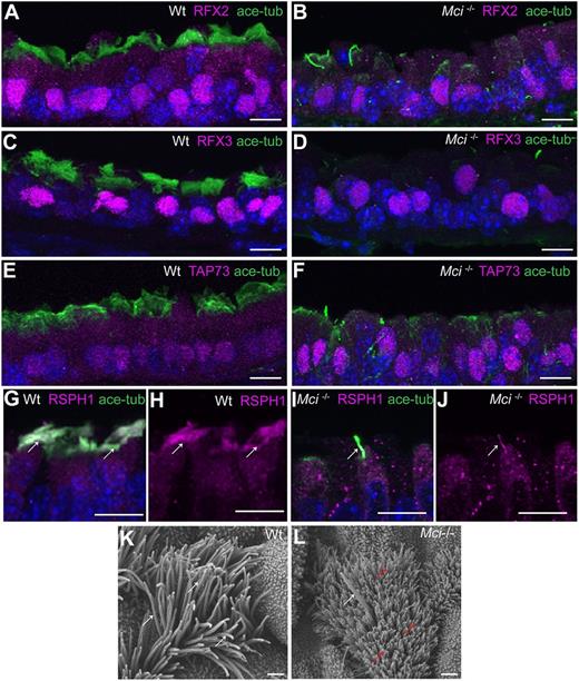
Fig. 2 Mci mutant MCC precursors express a suite of ciliary transcription factors and their single cilium localizes motile cilia-specific proteins. (A) Nuclear-localized RFX2 expression in MCCs of wild-type trachea. (B) Nuclear-localized RFX2 expression in monociliated cells of Mci mutant trachea. (C) Nuclear-localized RFX3 expression in MCCs of wild-type trachea. (D) Nuclear-localized RFX3 expression in monocilated cells of Mci mutant trachea. (E) Nuclear-localized TAP73 expression in MCCs of wild-type trachea. (F) Nuclear-localized TAP73 expression in monociliated cells of Mci mutant trachea. (G) RSPH1 colocalization with acetylated tubulin to MCC cilia of wild-type trachea (arrows). (H) RSPH1 localization to MCC cilia of wild-type trachea (arrows; display of only RPSH1 staining from G). (I) RSPH1 colocalization with acetylated tubulin to a single cilium of Mci mutant trachea (arrow). (J) RSPH1 localization to single cilium of Mci mutant trachea (arrow; display of only RSPH1 staining from I). (K) SEM analysis of a wild-type tracheal MCC showing multiple cilia (arrows). (L) SEM analysis of Mci mutant MCCs with a single cilium (white arrow). The microvilli, which are longer in the MCCs and normally remain obscured by the multiple cilia, are indicated (red arrows). One wild-type and one mutant trachea were scanned by SEM. The single-cilium phenotype of the Mci mutant trachea is representative of several fields of view scanned by SEM. In all preparations, cilia were stained using anti-acetylated tubulin antibodies (green) and nuclei with DAPI (blue). Scale bars: 10 μm in A-J; 5 µm in K,L.