
|
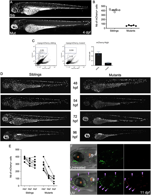
Fig. 1 In moonshine mutant zebrafish, primitive macrophages prematurely disappear between 54 hpf and 4 dpf. (A) mCherry+ macrophages in live 4 dpf Tg(mpeg1:mCherry-F) control sibling (top) and monNQ039 mutant (bottom) larvae; maximum projection, lateral view. (B) Quantification of mCherry+ macrophages in live Tg(mpeg1:mCherry-F) sibling (white dots) and monNQ039 mutant (black dots) larvae at 4 dpf (n=5 larvae per condition). Data are mean±s.e.m. (C) Quantification of mCherry+ macrophages from monNQ039/mpeg1:mCherry-F sibling (left plot) and mutant (right plot) larvae at 4 dpf by FACS. Plots provide representative data; graph provides data from a pool of two experiments (mean±s.e.m.). (D) mCherry+ macrophages in live Tg(mfap4:mCherry-F) zebrafish larvae at 48, 54, 72 and 96 hpf in control siblings (left) and monTB222 mutants (right); maximum projection. (E) Quantification of total mCherry+ macrophages in single monTB222/Tg(mfap4:mCherry-F) sibling and mutant larvae followed from 2 to 4 dpf (n=8 larvae per condition). (F) Head region at 11 dpf of Tg(mpeg1:Gal4;UAS:Kaede) larvae: untreated (top) or UV-photoconverted at 2 dpf (bottom); single confocal planes. Purple arrowheads indicate double-positive (i.e. photoconverted) macrophages. e, eye; E, ear.