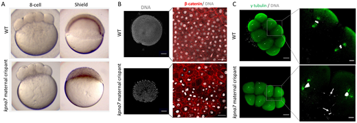
Fig. 4 Kpna7 is necessary for nuclear segregation during early development. (A) Images of live kpna7 maternal crispant and wild-type (WT) controls at the 8-cell stage (75 mpf) and shield stage (6 hpf). At the 8-cell stage, kpna7 maternal crispants appear to divide normally, but they stall at the sphere stage and fail to undergo epiboly. (B) Immunohistochemistry labeling of β-catenin and DAPI staining at 6 hpf showing that the kpna7 maternal crispant embryos exhibit nuclei of unequal sizes, including a subset of cells that entirely lack nuclei (blue asterisks). (C) Immunohistochemistry labeling of γ-tubulin and DAPI staining at 75 mpf, showing that kpna7 maternal crispant embryos display abnormal nuclear segregation (arrows) during cell cleavage. Scale bars: 100 μm (B,C, low magnification); 20 μm (B,C, high magnification).
Image
Figure Caption
Acknowledgments
This image is the copyrighted work of the attributed author or publisher, and
ZFIN has permission only to display this image to its users.
Additional permissions should be obtained from the applicable author or publisher of the image.
Full text @ Development

