
|
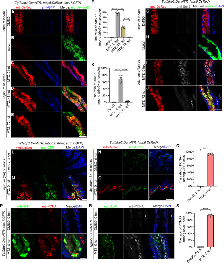
Fig. 6 Migrated ileal enterocytes in both larvae and adults express gut precursor markers (A–K) In the uninjured larvae, ileal enterocytes did not express sox17:GFP transgene and Sox9 (A and G). In the MTZ-injured larvae, the migrated DsRed+ ileal enterocytes in the jejunum expressed the sox17:GFP transgene (B and C) and Sox9 (H and I) at 0 hpt, followed by expression of Dendra2 at 48 hpt (D), finally downregulating sox17:GFP transgene (E) and Sox9 (J) at 72 hpt. (F) Graph showing quantification of ratio of Sox17+ among DsRed+ enterocytes (n = 6). (K) Graph showing quantification of ratio of Sox9+ among DsRed+ enterocytes (n = 6). Data are presented as mean ± SEM. ∗∗∗∗p < 0.0001, two-tailed unpaired t test. (L–O) Similarly, in MTZ-injured adults, the migrated DsRed+ ileal enterocytes in the S4 segment of jejunum expressed the sox17:GFP transgene (L and M) and Sox9 (N and O) at 0 hpt. (P–S) The sox17+ (P) and sox9+ (R) cells expressed PCNA. Graphs show ratio of PCNA+ cells among the sox17+ (Q) and sox9+ (S) cells. Data are presented as mean ± SEM. ∗∗∗∗p < 0.0001, two-tailed unpaired t test. Scale bars, 50 μm. See also Figure S7.