
|
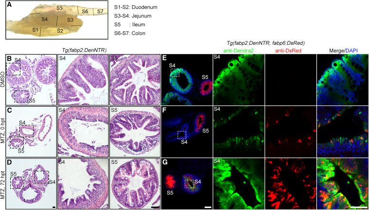
Fig. 5 Ileal enterocytes contribute to jejunal enterocyte regeneration in adult zebrafish (A) The adult zebrafish intestine is subdivided into seven segments (S1 to S7). (B–D) H&E staining shows that in contrast to the uninjured control (B), the jejunal villi in S4 collapsed at 0 hpt and underwent morphological reconstructing at 72 hpt, whereas the ileal villi in S5 remained unaffected (C and D). (E–G) In the uninjured Tg(fabp2:DenNTR; fabp6:dsRed) transgenic adults, DsRed+ enterocytes were present in S5 but not in S4 (E). After MTZ-induced injury at 0 hpt, the DsRed+ enterocytes were observed in S4 (F), which became more evident at 72 hpt (G). The dash-framed areas are enlarged in the panels on their right. Scale bars, 50 μm.