
|
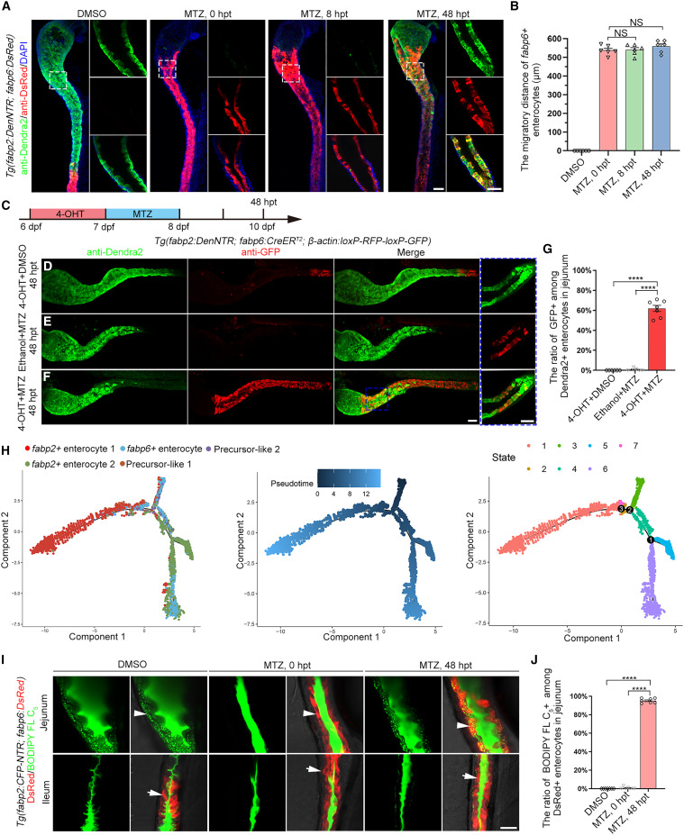
Fig. 4 Migrated ileal enterocytes transdifferentiate into jejunal enterocytes (A) Compared with the uninjured larvae under the Tg(fabp2:DenNTR; fabp6:dsRed) transgenic background, fabp6+ enterocytes migrated to jejunum at 0, 8, and 48 hpt. The migrated DsRed+ enterocytes in the injured jejunum expressed the fabp2:Dendra2 transgene at 48 hpt. The dash-framed areas are enlarged in the panels on their right. (B) Graph showing quantification of migratory distance (n = 6). Data are presented as mean ± SEM. Not significant (NS), two-tailed unpaired t test. (C–G) Stratagy of 4-OHT treatment (C). The Cre/loxP-mediated lineage tracing of ileal enterocytes. Uninjured larvae treated with 4-OHT served as the positive control, in which the ileal enterocytes were labeled with GFP (D). Negatively controlled by ethanol plus MTZ treatment (E), larvae treated with 4-OHT and MTZ exhibited GFP+Dendra2+ enterocytes in the injured jejunum (F). The dash-framed areas are enlarged in the right-hand panel. The statistics show that approximately 60% of the regenerated jejunal enterocytes were positive for GFP at 48 hpt (G, n = 7). Data are presented as mean ± SEM. ∗∗∗∗p < 0.0001, two-tailed unpaired t test. (H) A total of 8,234 cells were used for pseudotime trajectory of fabp2+ enterocyte, fabp6+ enterocyte, and precursor-like. The gradient from dark to light blue denotes the time increment. The seven states were identified by gene expression profiles in pseudotime. (I) BODIPY FL C5 green fluorescent droplets indicate lipid absorption. The enterocytes in the ileum are devoid of lipid uptake function (arrows). However, the ileal enterocytes migrated into the injured jejunum at 48 hpt and gained the function of lipid absorption (arrowheads). (J) Graph showing quantification of rate of BODIPY FL C5+ among DsRed+ enterocytes. Data are presented as mean ± SEM. ∗∗∗∗p < 0.0001, two-tailed unpaired t test. Scale bars, 50 μm. See also Figures S4 and S5.