
|
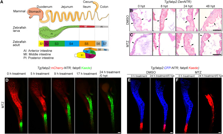
Fig. 1 Ileal enterocytes migrate to the extensively injured jejunum (A) The anterior, middle, and posterior intestine of zebrafish larvae correspond to mammalian duodenum plus jejunum, ileum, and colon, respectively. The S1-S2, S3-S4, S5, and S6-S7 intestinal segments of zebrafish adults are equivalent to mammalian duodenum, jejunum, ileum, and colon, respectively. (B and C) In contrast to the uninjured intestine (B), H&E staining showed that the intestinal villi collapsed at 0 hpt and 8 hpt after MTZ-induced injury, then rapidly recovered at 24 hpt and 48 hpt (C). The arrowheads indicate basement membrane, while the arrows indicate villus. (D) Live imaging showed that the Kaede-green+ ileal enterocytes quickly responded to the MTZ-induced jejunal injury and migrated into the jejunum. See Video S1. (E and F) Photoconversion of Kaede-green to Kaede-red showed that in contrast to the uninjured control (E), the Kaede-red+ ileal enterocytes migrated into jejunum in response to extensive jejunal enterocyte damage (F). “24 h treatment” is equivalent to 0 hpt. Scale bars, 50 μm. See also Figures S1 and S2.