
|
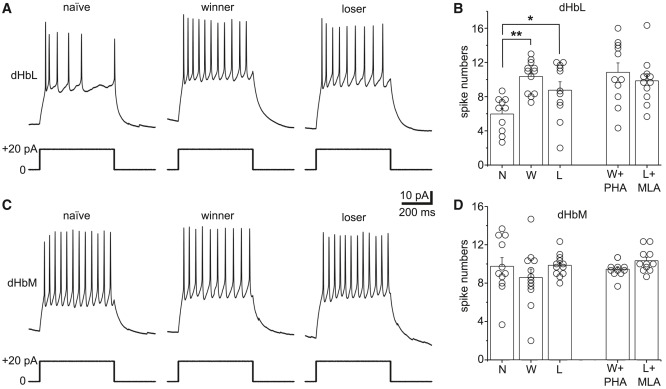
Fig. 7 The excitability of the Hb does not differ between winners and losers (A) Representative spiking activities (top) induced by current injection (bottom) in a dHbL neuron from naive (left), winner (middle), or loser (right) fish. (B) Comparison of the numbers of recorded spikes in dHbL neurons among naive (N, n = 10 cells from 3 animals), winner (W, n = 12 cells from 3 animals), loser (L, n = 11 cells from 3 animals), winner + PHA (W + PHA, n = 12 cells from 3 animals), and loser + MLA (L + MLA, n = 12 cells from 3 animals) groups. (C) Representative spiking activities (top) induced by current injection (bottom) in a dHbM neuron from naive (left), winner (middle), or loser (right) fish. (D) Comparison of the numbers of recorded spikes in dHbM neurons among naive (N, n = 11 cells from 3 animals), winner (W, n = 12 cells from 3 animals), loser (L, n = 12 cells from 3 animals), winner + PHA (W + PHA, n = 11 cells from 3 animals), and loser + MLA (L + MLA, n = 11 cells from 3 animals) groups. Values are presented as mean ± SEM. Statistical significance was defined as ∗p < 0.05; ∗∗p < 0.01. Scale bars for (A) and (C), 10 pA, 200 ms.