
|
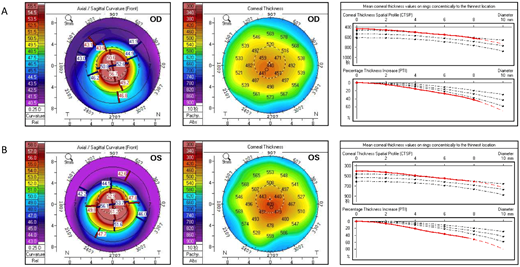
Fig. 1 Clinical ocular features of the ZNF469 mutant patient. (A) Pentacam refractive maps and corneal thickness map of the right eye (OD). Thinnest corneal thickness = 435 µm, Kmax = 64.9 D. (B) Pentacam refractive maps and corneal thickness map of the left eye (OS). Thinnest corneal thickness = 406 µm, Kmax = 70.4 D. Pupil center is indicated by a plus sign (+), the pachy apex is marked with a filled circle (•), and the thinnest location is indicated by an open circle (○). Three black dotted lines in the corneal thickness map indicate the distribution of healthy people, and the red line indicates the fitting curve of the patient.