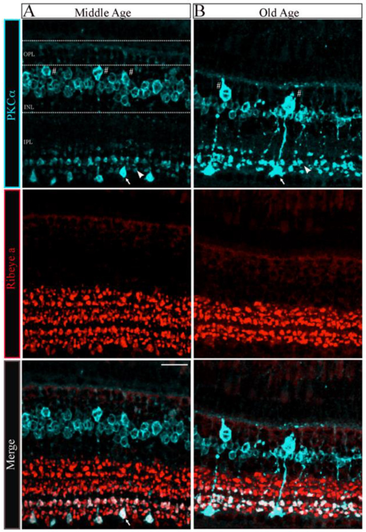
The morphology of bipolar cell ribbon synapses in the retinal IPL was altered in older-aged zebrafish. (A,B) Transverse retinal sections from middle-aged ((A), left panels; n = 2~4 sections in four retinas from two fish) and old-aged ((B), right panels; n = 2~4 sections in four retinas from two fish) zebrafish were double immunostained with fluorescently labeled antibodies specific for the rod bipolar cell marker PKCα (cyan; top panels) or ribeye a (red; middle panels); also shown is the overlay of PKCα and ribeye a labeling (merge; bottom). Maximal intensity projections are shown, and the relative positions of the INL and IPL are indicated. Scale bar, 20 µm. PKCα, protein kinase C alpha; OPL, outer plexiform layer; INL, inner nuclear layer; IPL, inner plexiform layer. Larger (arrow) and smaller (arrowhead) are indicated in PKCα ribbon terminal morphologies in MA and OA. # Denotes the pear-shaped soma characteristics of Mb1 rod bipolar cells with larger terminals.
Acknowledgments
This image is the copyrighted work of the attributed author or publisher, and
ZFIN has permission only to display this image to its users.
Additional permissions should be obtained from the applicable author or publisher of the image.
Full text @ Cells
Your Input Welcome
Thank you for submitting comments. Your input has been emailed to ZFIN curators who may contact you if
additional information is required.
Oops. Something went wrong. Please try again later.