
IMAGE
Fig. 5
- ID
- ZDB-IMAGE-230912-11
- Publication
- Zhang et al., 2023 - Knockout of miR-184 in zebrafish leads to ocular abnormalities by elevating p21 levels
- All Figures
- Figures for Zhang et al., 2023
Image
|
Figure Caption
Fig. 5
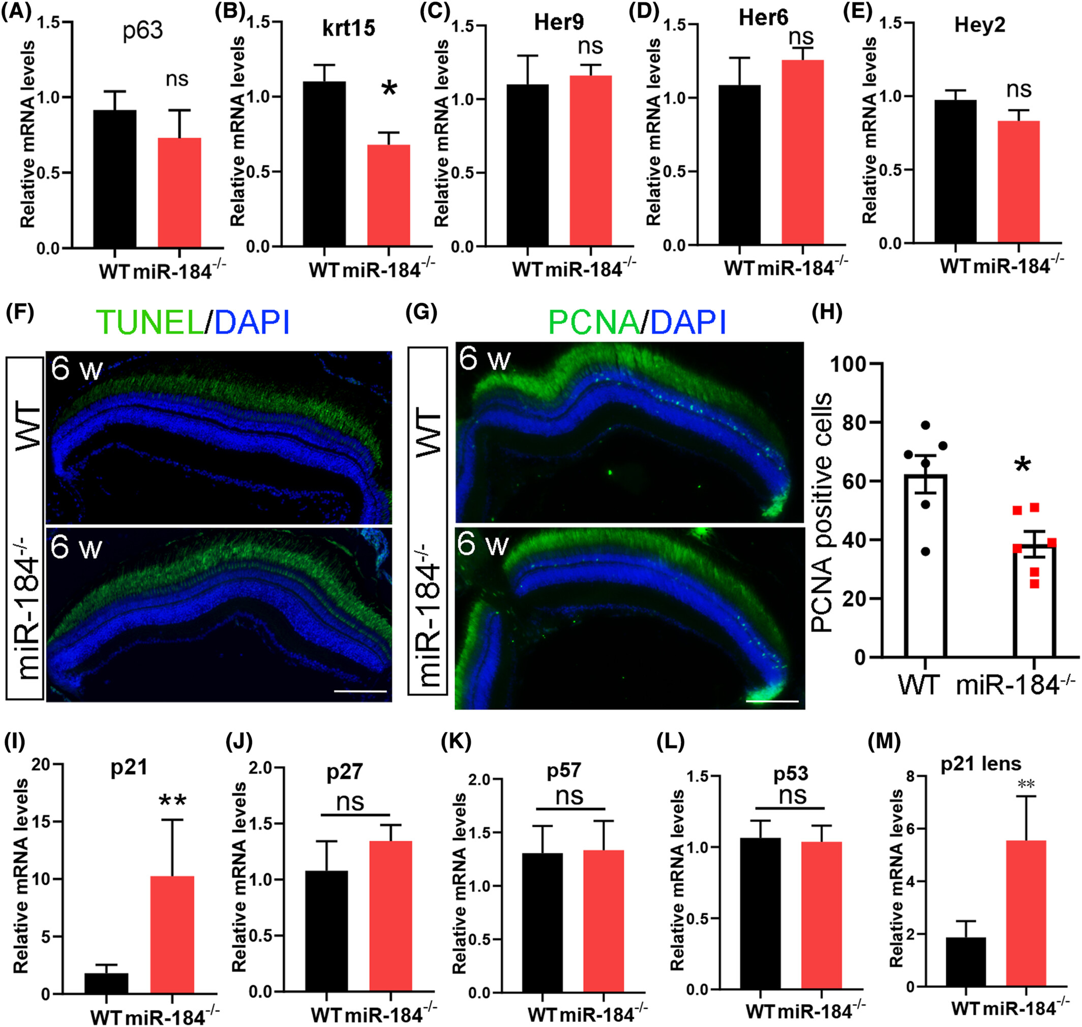
Depletion of miR-184 resulted in aberrant cell proliferation and elevated p21 levels. (A–E) The mRNA levels of p63, krt15, Her9, Her6, and Hey2 were analyzed in 3-month-old WT and miR-184−/− optic cups (eye tissue without lens). Student t-test was used for statistical analysis, n = 3. p < .05. (F) The TUNEL signals were detected in 1.5-month-old WT and miR-184−/− retinas. The nuclei were stained with DAPI. Bar, 100 μm. (G) The PCNA signals were detected in 1.5-month-old WT and miR-184−/− retinas. The nuclei were stained with DAPI. Bar, 100 μm. (H) The number of PCNA-positive cells in (B) were counted. Student t-test was used for statistical analysis, n = 6. *p < .05. (I–L) The mRNA levels of p21, p27, p57, and p53 were analyzed in 3-month-old WT and miR-184−/− optic cups (eye tissue without lens). Student t-test was used for statistical analysis, n = 3. **p < .01. (M) The p21 mRNA levels were analyzed in 3-month-old WT and miR-184−/− lenses. Student t-test was used for statistical analysis, n = 3. **p < .01.
Acknowledgments
This image is the copyrighted work of the attributed author or publisher, and
ZFIN has permission only to display this image to its users.
Additional permissions should be obtained from the applicable author or publisher of the image.
Full text @ FASEB J.