
Image
|
Figure Caption
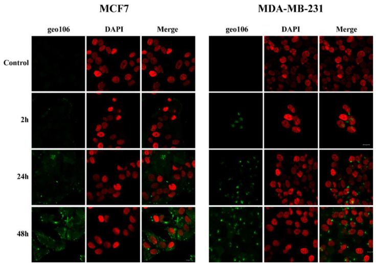
Fig. 7
Confocal microscopy images of MCF7 and MDA-MB-231 cells after incubation with 5 μM of geo106 for 2, 24 and 48 h, respectively. Cells were fixed with 4% paraformaldehyde. DNA was stained with DAPI (pseudo color red). Cells were visualized with a Zeiss LSM 780 confocal microscope using the Zen 2011 software.
Acknowledgments
This image is the copyrighted work of the attributed author or publisher, and
ZFIN has permission only to display this image to its users.
Additional permissions should be obtained from the applicable author or publisher of the image.
Full text @ Pharmaceutics