Image
Figure Caption

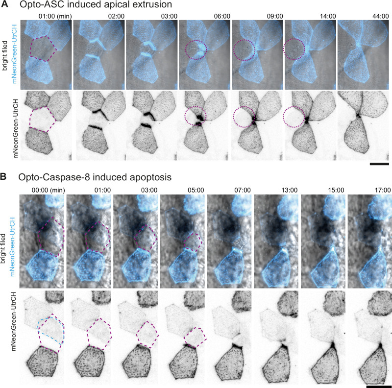
Figure 6—figure supplement 2. Actin rearrangement in periderm cells near dying cells.

(A, B) Actin, labelled by mNeonGreen-Utr-CH (cyan in top row and black in the bottom row) is expressed in a mosaic pattern. As the surrounding cells close the gap over the dying cells, actin accumulates at the lateral membranes which are in contact with the dying cell. (A) Response of neighboring periderm cells to Opto-ASC induced apical extrusion. Opto-ASC is induced in the periderm cell marked in purple dotted line. (B) Response of neighboring cells to apoptosis induced by Opto-Caspase 8 in the periderm cell marked in purple. This cell does not express mNeonGreen-Utr-CH, but the signal comes from an underlying basal cell (outlined in blue).

Acknowledgments
This image is the copyrighted work of the attributed author or publisher, and ZFIN has permission only to display this image to its users. Additional permissions should be obtained from the applicable author or publisher of the image. Full text @ Elife