
IMAGE
Figure 3—figure supplement 1.
- ID
- ZDB-IMAGE-230710-22
- Source
- Figures for Hasel de Carvalho et al., 2023
Image
|
Figure Caption
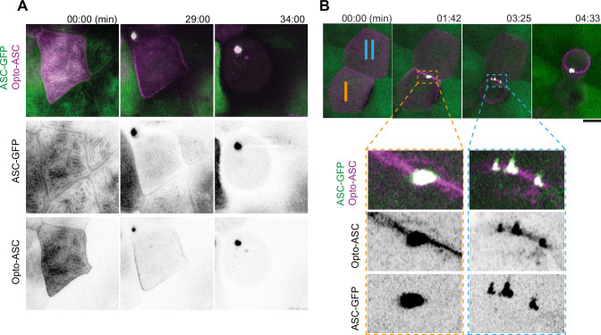
Figure 3—figure supplement 1. Time lapse of speck formation induced by Opto-ASC (magenta) and recruitment of endogenous ASC (green, ubiquitous) in a periderm cells. Scale is 10 µm. (B) Two neighboring cells (I and II) forming either a single speck (cell I) or multiple specks (cell II) along the cell membrane. The specks coalesce as the cell shrinks. Scale is 2 µm.
Acknowledgments
This image is the copyrighted work of the attributed author or publisher, and
ZFIN has permission only to display this image to its users.
Additional permissions should be obtained from the applicable author or publisher of the image.
Full text @ Elife