
Fig. 2
- ID
- ZDB-IMAGE-230617-9
- Publication
- Sun et al., 2023 - hapln1a+ cells guide coronary growth during heart morphogenesis and regeneration
- All Figures
- Figures for Sun et al., 2023
|
Fig. 2
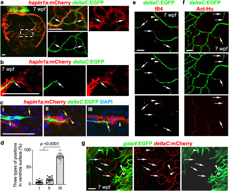
a Whole-mount view of hapln1a+ cells and deltaC:EGFP+ coronary endothelial cells on hapln1a:mCherry-NTR;deltaC:EGFP ventricular surface (opposite the atrioventricular junction) at 7 weeks post-fertilization (wpf). The boxed area is enlarged. Arrows indicate deltaC:EGFP+ cells lining with hapln1a+ cells. n = 50 animals. Scale bar, 100 µm. b Representative images of hapln1a+ cells located ahead of deltaC:EGFP+ cells in the juvenile ventricular surface. Arrows indicate coronary tips. Total 50 animals have been examined from 3 independent experiments with similar results. Scale bar, 50 µm. c The three position types of hapln1a+ cells according to their localization with coronary sprouts in the juvenile heart surface: hapln1a+ cells behind (I), parallel (II), and leading (III) deltaC+ cells. Arrows, front of deltaC+ cells. Arrowheads, front of hapln1a+ cells. Scale bar, 50 µm. d Quantification of the three types of positions from experiments in c. I, 6%; II, 12%; III, 82%. n = 23 animals. Mann–Whitney rank-sum test (Two-sided). Data are presented as mean values ± SD. Source data are provided as a Source Data file. e Growing deltaC+ cell extensions and macrophages (IB4+) in juvenile hearts. Arrowheads represent IB4+ macrophages flanking coronary vessels. Arrows represent deltaC+ vessel extensions. n = 8 animals. Scale bar, 50 µm. f Growing deltaC+ cell extensions and AcT-Hu+ nerves in juvenile hearts. Arrows represent EGFP+ vessel extensions. n = 5 animals. Scale bar, 50 µm. g Growing deltaC+ cell extensions and gata4+ CMs in juvenile hearts. Arrows represent deltaC+ vessel extensions outside gata4+ CM area. Arrowheads represent deltaC+ vessel extensions embedded in gata4+ CM area. n = 8 animals. Scale bar, 50 µm.