
Fig. 5
- ID
- ZDB-IMAGE-230617-12
- Publication
- Sun et al., 2023 - hapln1a+ cells guide coronary growth during heart morphogenesis and regeneration
- All Figures
- Figures for Sun et al., 2023
|
Fig. 5
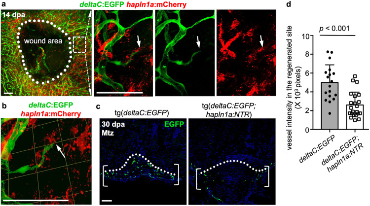
a Whole-mount view of coronary vessels and hapln1a+ cells in the wound area of adult injured deltaC:EGFP;hapln1a:mCherry animals. White dashed box is enlarged in single confocal slices. Arrows label the coronary tips. White dashed line circles the closing wound. n = 10 animals. Scale bars, 50 µm. b 3-D visualization of coronary sprouts and hapln1a+ cells in the regenerating area in experiment (a). Arrow represents the coronary tip lagging behind hapln1a+ cells. Scale bars, 50 µm. c Section views of coronary vessels in the wounded area in adult injured deltaC:EGFP;hapln1a:mCherry animals and deltaC:EGFP siblings, with Mtz treatment by 30 dpa. Brackets represent the regenerated area. Scale bars, 50 µm. d Quantification of EGFP+ pixels in the regenerating site from (c). n = 18 deltaC:EGFP and n = 22 deltaC:EGFP;hapln1a:mCherry animals were used. The experiments were repeated at least once. Mann–Whitney rank-sum test (Two-sided). Data are presented as mean values ± SD. Source data are provided as a Source Data file.