IMAGE

Fig. 1.

ID
ZDB-IMAGE-230609-33
Source
Figures for Maili et al., 2023
Image
Figure Caption

Fig. 1.

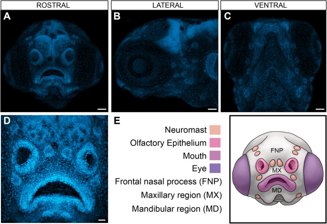
Craniofacial features visualized in frontal/rostral mounts by DAPI staining and confocal microscopy. (A-D) Rostral (A,D), lateral (B) and ventral (C) views of developing zebrafish larvae at 5 days post-fertilization (dpf). (E) Schematic representing the facial anatomical structures visualized in the rostral view, including neuromasts, olfactory placodes, eyes, mouth, frontonasal region (FNP), maxillary region (MX) and mandibular region (MD). Scale bars: 50 µm (A-C), 20 µm (D).

Acknowledgments
This image is the copyrighted work of the attributed author or publisher, and ZFIN has permission only to display this image to its users. Additional permissions should be obtained from the applicable author or publisher of the image. Full text @ Dis. Model. Mech.