
Image
|
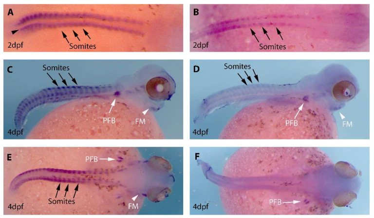
Figure Caption
Figure 3
Expression analysis of MyoD1 and MyoD2 in the developing embryos of Oreochromis (Alcolapia) alcalica. (A,C,E) Lateral and dorsal views of O. alcalica embryos analysed by in situ hybridisation for MyoD1 and (B,D,F) for MyoD2. The number of days post fertilisation [dpf] is indicated for each image. Arrows show somites, white arrowheads indicate facial muscle (FM) and white arrows indicate developing pectoral fin bud (PFB). Black dots around the yolk and on the body are chromatophores (pigment cells).
Acknowledgments
This image is the copyrighted work of the attributed author or publisher, and
ZFIN has permission only to display this image to its users.
Additional permissions should be obtained from the applicable author or publisher of the image.
Full text @ J Dev Biol