
|
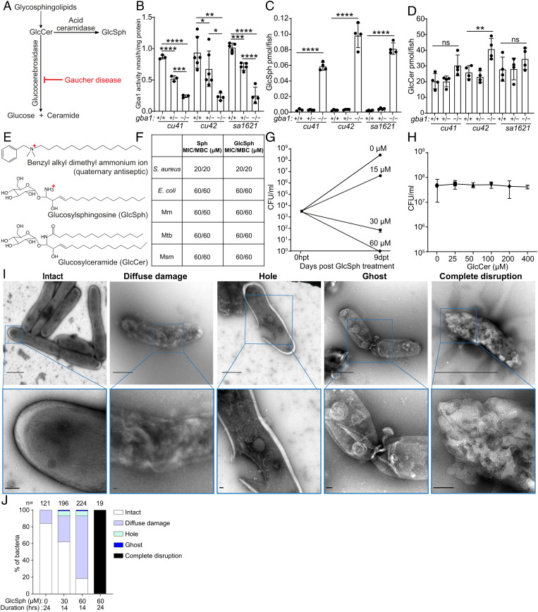
Fig. 4.
Glucosylsphingosine is bactericidal in vitro. (A) Representation of the pathway leading to glucosylceramide and glucosylsphingosine accumulation in Gaucher disease. GlcCer, glucosylceramide. GlcSph, glucosylsphingosine. (B) Glucocerebrosidase enzyme activity (nmol/mg) in 5 dpf gba1 cu41, cu42, and sa1621 mutants and their wild-type siblings. Mean ± SD; *P < 0.05; **P < 0.01; ***P < 0.001; ****P < 0.0001 (one-way ANOVA with Tukey’s posttest). (C) GlcSph (pmol/fish) in 5 dpf gba1 cu41, cu42, and sa1621 mutants and their wild-type siblings. Mean ± S.D; ****P < 0.0001 (one-way ANOVA with Tukey’s posttest). Representative of two independent experiments. (D) GlcCer (pmol/fish) in 5 dpf gba1 cu41, cu42, and sa1621 mutants and their wild-type siblings. Mean ± SD; ns, not significant; **P < 0.01 (one-way ANOVA with Tukey’s posttest). Representative of two independent experiments. (E) Chemical structures of benzyl alkyl dimethyl ammonium ion, GlcSph, and GlcCer. (F) MIC/MBC table of Staphylococcus aureus, Escherichia coli, Mm, Mtb, and M. smegmatis treated with Sph and GlcSph. Representative of one to two independent experiments performed in duplicate. (G) Mm killing by GlcSph. Mean CFU/mL; vertical bars, upper and lower values of the two technical replicates. (H) Mean Mm (CFU/mL) after incubation with increasing GlcCer concentrations for 9 d. Vertical bars, upper and lower values of the two technical replicates (starting concentration, 2.8 × 10³ CFU/mL). Representative of two independent experiments. (I) Negative stain TEM images of Msm treated with GlcSph, representative of the types of damage seen. [Scale bars, 1 μm (Top) and 100 nm (Bottom).] (J) Quantification of the types of damage seen in (H) for various GlcSph concentrations.