
|
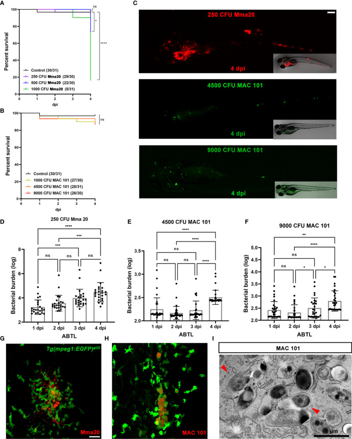
Figure 1
Characterization of M. avium infection in zebrafish larvae compared to M. marinum infection. (A, B) Percent of survival curves for ABTL zebrafish larvae infected with a series of doses M. marinum Mma20 or M. avium MAC 101. ABTL zebrafish larva infected with mCherry-labeled M. marinum Mma20 at a dose of ~250 CFU and infected with wasabi-labeled M. avium MAC 101 at a dose of ~4500 CFU or 9000 CFU by caudal vein infection at 28 hpf. (C) Representative images for the bacterial burden quantification were taken at 4 dpi. Scale bar: 50 µm. (D) Bacterial burden quantification of ABTL zebrafish larvae upon ~250 CFU Mma20 infection. (E) Bacterial burden quantification of ABTL zebrafish larvae upon ~4500 CFU MAC 101 infection. (F) Bacterial burden quantification of ABTL zebrafish larvae upon ~9000 CFU MAC 101 infection. (G, H) Representative CLSM images of Tg(mpeg1: EGFP)gl22 zebrafish larvae infected with mCherry-labeled Mma20 strain (G) or DsRed-labeled MAC 101 (H). Tg(mpeg1: EGFP)gl22 embryos were infected ~250 CFU Mma20 mCherry strain or ~4500 CFU MAC 101 DsRed strain at 28 hpf. CLSM images were taken for the 4 dpi infected larvae by using 40 times magnification lens (oil immersion, N.A. 1.3). Scale bar: 50 µm. (I) TEM pictures showing a sagittal section through MAC 101 in wild type zebrafish larva. Red arrows represent the bacteria inside of a phagocyte. Scale bar: 1 µm. In (A, B) data were collected from three pools of zebrafish larvae. In (D, E, and F) data (mean ± SD) were combined from three pools of zebrafish larvae. Statistical significance of differences was determined by using one-way ANOVA with Tukey’s multiple comparison test as a post-hoc test. *P < 0.05, **P < 0.01, ***P < 0.001, ****P < 0.0001. Sample size (n): 24, 24, 23, 24 (D), 31, 33, 31, 30 (E), 30, 29, 27, 29 (F).