
Fig. 2.
|
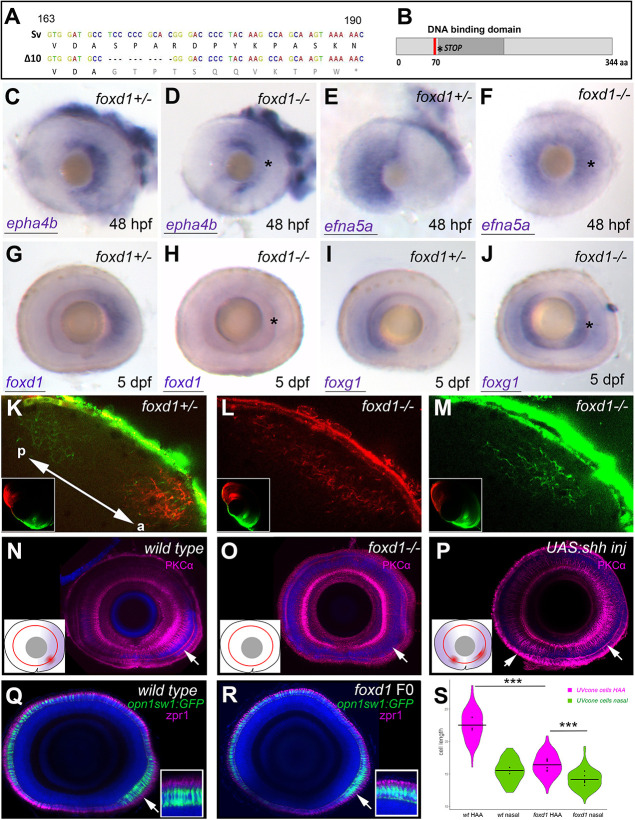
Fig. 2. Loss of foxd1 results in retinae with expanded nasal and reduced temporal character. (A) foxd1 sequence comparison between nucleotides 163 and 190 of the open reading frame, highlighting the 10 bp deletion (Δ10, bottom row) in the foxd1cbm16 mutant. (B) Diagrammatic representation of the Foxd1 protein, depicting (in red) the deleted region in the cbm16 allele. The asterisks in A and B mark the position at which a newly generated stop codon is present. (C-J) Lateral views with nasal towards the left of 48 hpf (C-F) and 5 dpf (G-J) retinae, showing expression of nasal or temporal markers in foxd1−/− mutants and siblings. The markers assessed and the genotypes of the retinae are given in the bottom left and top right corners, respectively. Asterisks in D,F,H,J highlight altered expression in foxd1 mutants. Twenty-five to 30 embryos were processed together per marker. foxd1−/− embryos were recovered at the expected mendelian proportions and the phenotypes observed were fully penetrant. Genotype of imaged embryos was confirmed by molecular genotyping. (K-M) Dorsal views of the tectum of foxd1−/− mutant (L,M; n=12) and sibling (K; n=10) embryos, with nasal RGC arbours labelled using DiO (green) and temporal RGC arbours using DiI (red). Insets in K-M show dorsal views of the corresponding eye. Tectum and eye orientation in K-M are indicated by the double-headed arrow in K (a, anterior; p, posterior). (N-P) PKCα immunostaining (magenta) in lateral views with nasal towards the left of 8 dpf wild-type (N), foxd1−/− mutant (O) and tg{rx3:Gal4};UAS:Shh (P) eyes. Nuclei are counterstained using DAPI (blue). Insets in N-P are schematics of the eyes in the corresponding panels, highlighting the extent of temporal character (purple), as assessed by gene expression, and the areas enriched with PKCα (red). Arrows indicate the position of PKCα enrichment. (Q,R) Sagittal sections across wild-type (Q) and foxd1 crispant (R) eyes with nuclei stained using DAPI (blue), the outer photoreceptor segments stained using anti-Zpr1 antibodies (magenta) and UV cones expressing GFP [green, Tg(opn1sw1:GFP)] in 8 dpf larvae. (R) An example of a mild phenotype in comparison with other embryos with more severely reduced HAA labelling. (S) Violin plots showing differences in cone cell length between cells located in the HAA and nasal retina in wild type (n=3 eyes) and foxd1 crispants (n=6 eyes). Horizontal bars show the mean cell length for each condition; dots indicate the mean cell length for each individual eye (***P<0.001; one-way ANOVA) Scale bars: 100 µm.