
|
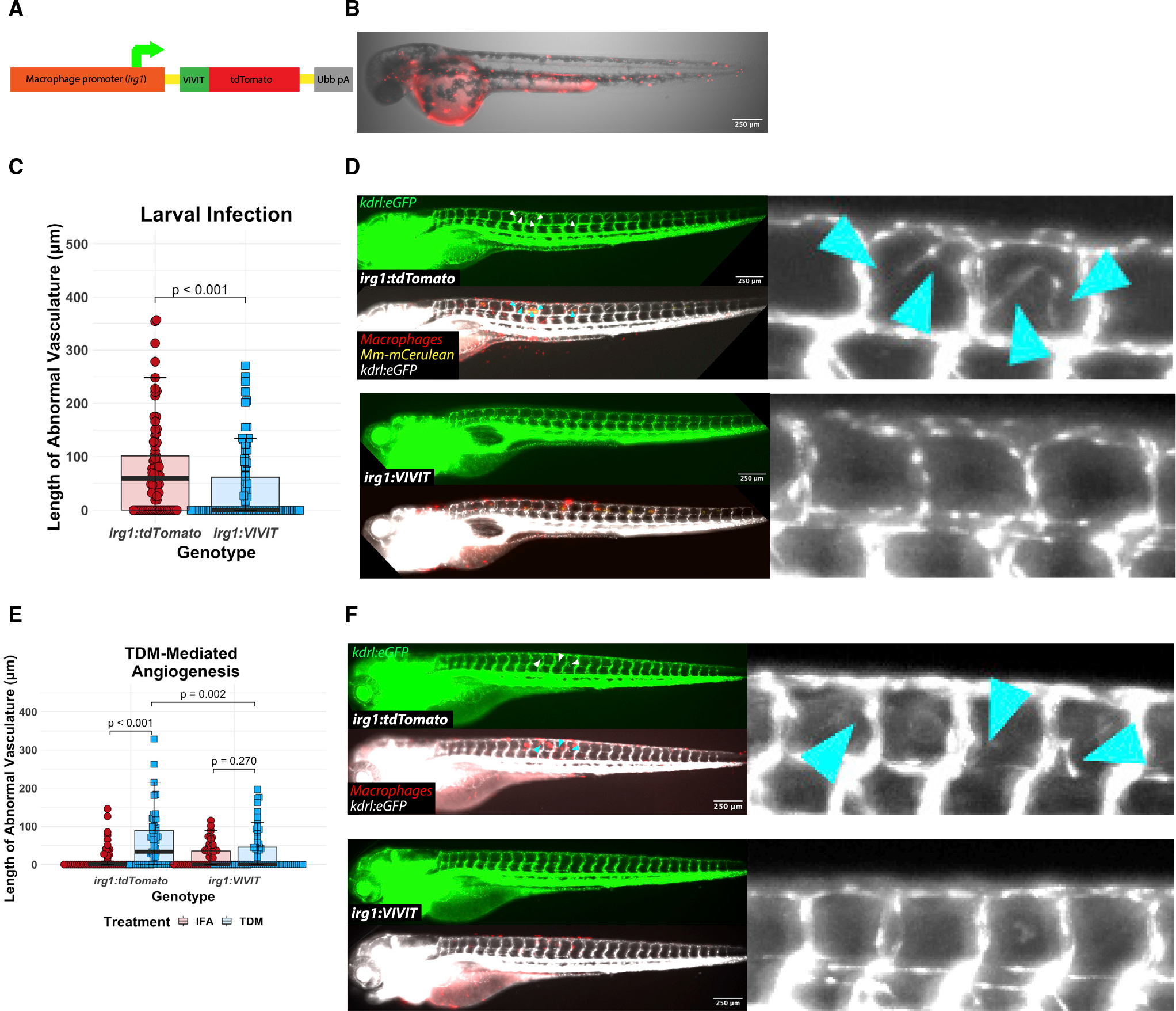
Figure 4. Macrophage-specific inhibition of NFAT signaling reduces angiogenesis in vivo during mycobacterial infection and in response to purified TDM
(A) Diagram of the Tg(irg1:VIVIT-tdTomatoxt39) line. The VIVIT peptide is directly conjugated to the tdTomato fluorescent protein and expression is driven by a −4.6 kb macrophage-specific irg1 promoter.
(B) Representative 2 day post fertilization larva showing macrophage-specific tdTomato+ expression throughout the larva. Note background expression in the yolk. Scale bar, 250 μm.
(C) Quantitation of angiogenesis during Mm infection of irg1:VIVIT-tdTomato or irg1:tdTomato larvae. irg1:VIVIT larvae display a statistically significant reduction in the degree of angiogenesis induced by infection at 4 dpi compared with irg1:tdTomato larvae. Statistics from Wilcoxon ranked-sign test. Representative of three independent biological replicates. Additional replicates are shown in Figures S2G and S2H; n = 92 tdTomato, 98 VIVIT.
(D) Representative images of irg1:tdTomato and irg1:VIVIT-tdTomato larvae at 4 dpi. VIVIT-expressing larvae display reduced neovascular elaboration compared with tdTomato-only controls.
(E) Quantitation of TDM-induced angiogenesis in irg1:VIVIT-tdTomato larvae compared with irg1:tdTomato larvae. Fish were injected with either TDM emulsified in IFA or IFA alone. Statistics were conducted by Dunn’s Kruskal-Wallis multiple comparisons test with Benjamini-Hochberg adjustment. Representative of three independent biological replicates. Additional replicates provided in Figures S2I and S2J; n = 59 tdTomato/IFA, 69 tdTomato/TDM, 62 VIVIT/IFA, 71 VIVIT/TDM.
(F) Representative images of TDM-injected larvae from either irg1:VIVIT-tdTomato or irg1:tdTomato animals. The irg1:VIVIT-tdTomato condition displays a reduction in angiogenesis to the level of background while irg1:tdTomato fish induce a robust angiogenic response. Arrowheads indicate regions of neovascularization.