
Image
|
Figure Caption
Fig. 6
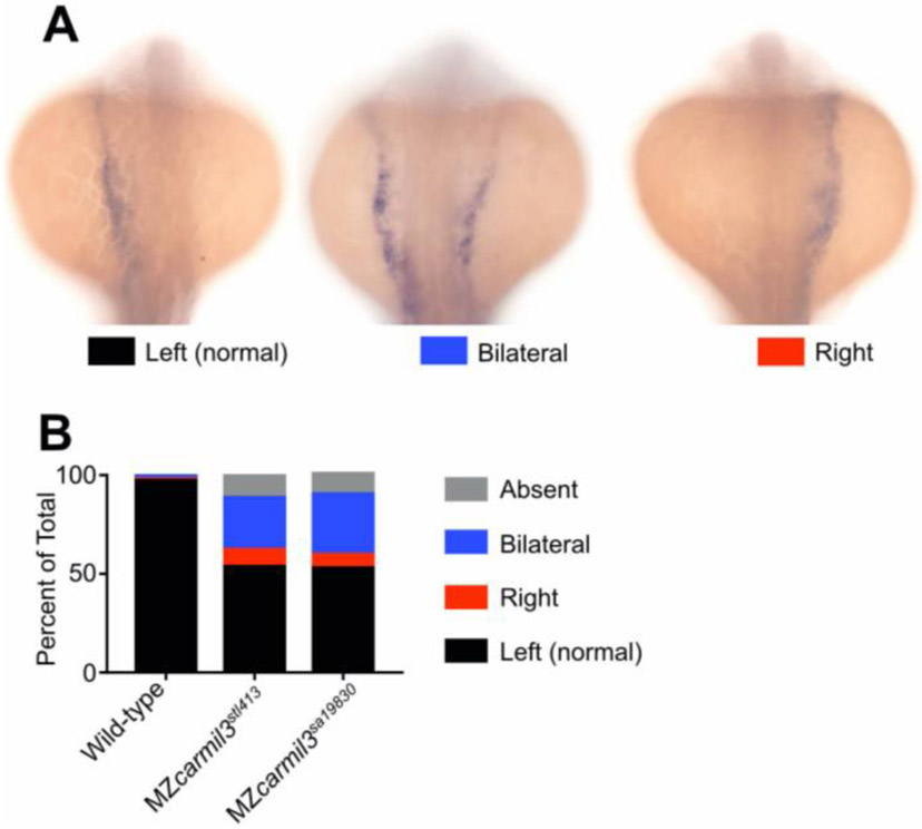
Patterns of spaw staining distribution at the 18-20 somite stage in carmil3 mutant embryos compared with WT embryos. (A) Representative images illustrate observed patterns of spaw staining, which is purple. (B) Percentage of spaw staining patterns, comparing WT embryos with embryos of two different carmil3 alleles, MZcarmil3stl413 and MZcarmil3sa19830. The color scheme is indicated in panel A, below the images. In cases scored as “absent,” no staining was observed. Values are listed in Table I.
Figure Data
Acknowledgments
This image is the copyrighted work of the attributed author or publisher, and
ZFIN has permission only to display this image to its users.
Additional permissions should be obtained from the applicable author or publisher of the image.
Reprinted from Developmental Biology, 481, Stark, B.C., Gao, Y., Sepich, D.S., Belk, L., Culver, M.A., Hu, B., Mekel, M., Ferris, W., Shin, J., Solnica-Krezel, L., Lin, F., Cooper, J.A., CARMIL3 is important for cell migration and morphogenesis during early development in zebrafish, 148-159, Copyright (2021) with permission from Elsevier. Full text @ Dev. Biol.