
|
Fig. 1
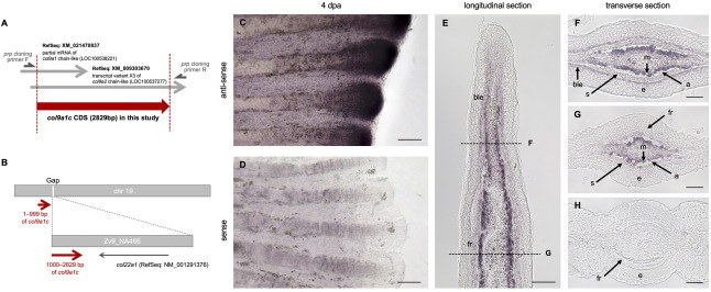
Fig. 1. Identification of col9a1c, newly isolated in this study. (A) Schematic diagram of the region of the isolated cDNA in this study (red arrow). This cDNA was named col9a1c (accession number: LC546948). (B) Schematic diagram of the position of the col9a1c in the database. Although the front part (1–999 bp) and the latter part (1000–2829 bp) are now on two different scaffolds, we inferred the unknown scaffold (Zv9_NA466) locates in the gap region of the chromosome (chr) 19 with considering the orientation of col9a1c gene. (C, D) Whole-mount in situ hybridization (ISH) of col9a1c for regenerated fins at 4 days post-amputation (dpa). Stronger expression was observed in the fin-tip. (E–H) ISH on cryo-sections of longitudinal (E) and transverse (F to H) at 4 dpa. The dot lines in E indicate the approximate positions of the transverse sections shown in F and G, respectively. At more distal fin-tip, stronger expression of col9a1c was observed in mesenchymal cells and the expression was also detected in basal layer of epidermis (F). More proximal region, the expression was mainly and strongly detected in mesenchymal cells (G). The expression was not observed in the proximal part of the regenerated fin, where thick fin-ray was present (H). ble: basal layer of epidermis, fr: fin-ray, s: scleroblast, e: epidermis, m: mesenchymal cells, a: actinotrichia. Scale bars: 100 μm in C, D; 50 μm in E; 25 μm in F–H.
Reprinted from Developmental Biology, 481, Nakagawa, H., Kuroda, J., Aramaki, T., Kondo, S., Mechanical role of actinotrichia in shaping the caudal fin of zebrafish, 52-63, Copyright (2021) with permission from Elsevier. Full text @ Dev. Biol.