
|
Fig. 5
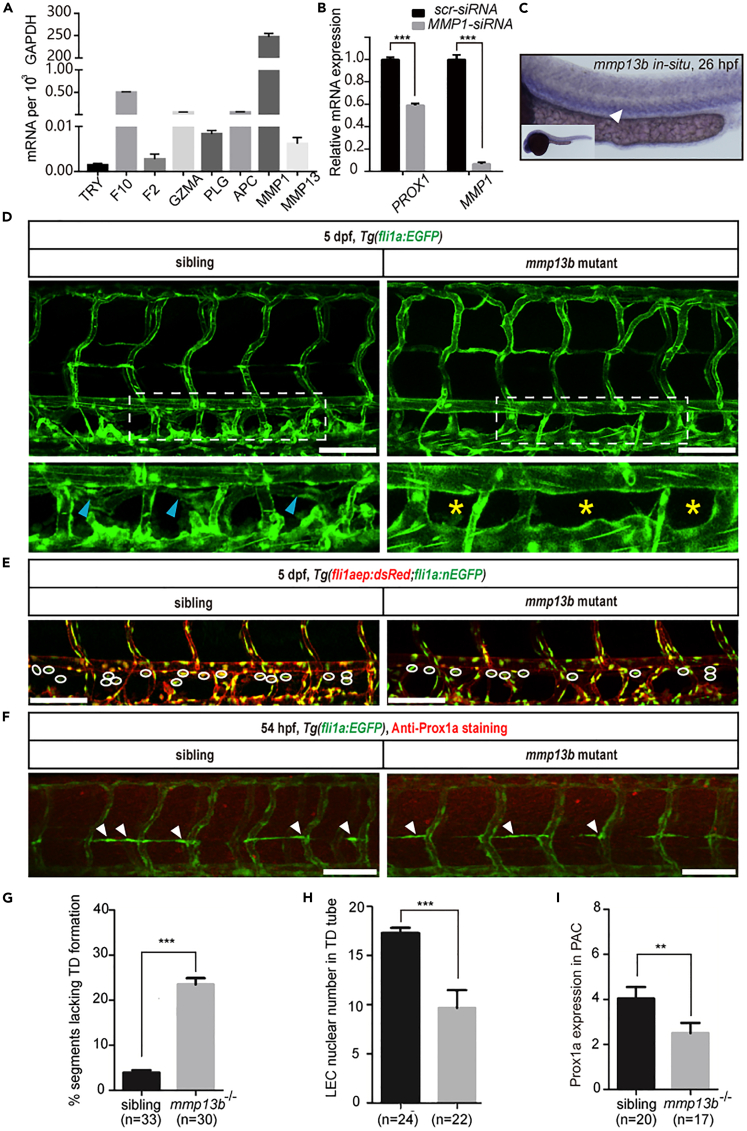
mmp13b mutant mimics the lymphatic phenotypes observed in the par1 mutant
(A) Relative mRNA expression of different PAR1 proteases in HDLECs.
(B) Relative mRNA expression of PROX1 and MMP1 in HDLECs after ctr-siRNA (control) and MMP1-siRNA transfection.
(C) WISH of mmp13b gene expression in zebrafish at 26 hpf. The white arrowhead indicates the expression of the mmp13b gene in the PCV area.
(D) Confocal images showing TD formation in Tg(fli1a:EGFP) siblings and mmp13b homozygous mutants at 5 dpf. Blue arrowheads indicate TD formation in each somite; yellow asterisks represent the absence of TD formation in each somite; DA and PCV areas are denoted. Scale bars: 100 μm.
(E) Confocal images showing LECs nuclear numbers in the TD tube of siblings and mmp13b homozygous mutants in the Tg(fli1aep: dsRed;fli1:nEGFP) line at 5 dpf. White circles indicate the presence of LECs nuclear numbers in the TD tube. Scale bars: 100 μm.
(F) Immunostaining of Prox1a of siblings and mmp13b homozygous mutants in the Tg(fli1a:EGFP) line at 54 hpf. White arrowheads indicate Prox1a-positive staining in the PLs. Scale bars: 100 μm.
(G) Percentage of somites lacking TD formation in siblings (n = 33 embryos) and mmp13b homozygous mutants (n = 30 embryos); 6 somites/embryos were used for quantification.
(H) LEC nuclear numbers in the TD tube of siblings (n = 24 embryos) and mmp13b homozygous mutants (n = 22 embryos); 6 somites/embryos were used for quantification.
(I) Prox1a-positive PLs in siblings (n = 20 embryos) and mmp13b homozygous mutants (n = 17 embryos) at 54 hpf; 7 somites/embryos were used for quantification. In (G–I), values represent means ± SEMs. ∗p ≤ 0.01, ∗∗p ≤ 0.001 in Student's t test. See also Figures S5 and S6.