
|
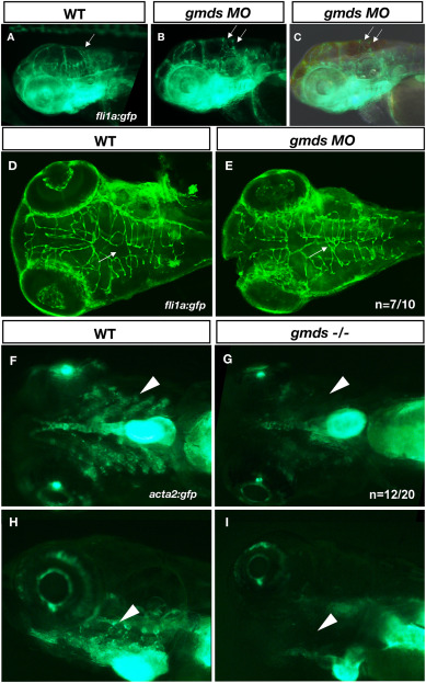
Fig. 4 Analysis of endothelial cell organization and smooth muscle cell number due to loss of gmds. In gmds morphant embryos, ectopic branches of cerebral vessels are observed at 3 dpf (B, C, E white arrows) when compared to control injected embryos (A, D). Panel C shows merged gfp and brightfield images to show the location of cerebral hemorrhage (hindbrain ventricle) near the site of an ectopic branch as highlighted by fli1a:gfp expression. gmds mutant embryos showed decreased smooth muscle actin cells in the bulbus arteriosus and pharyngeal arch arteries (G ventral view, I, lateral view) compared to wildype siblings (F, ventral view, H, lateral view) at 5 dpf. Embryos were deemed wildtype (+/+ or +/−) based on a lack of hemorrhage and curly tail, or mutant based on the presence cerebral hemorrhage and curly tail.
Reprinted from Developmental Biology, 480, Fowler, G., French, D., Rose, A., Squires, P., Anecito da Silva, C., Ohata, S., Okamoto, H., French, C.R., Protein fucosylation is required for Notch dependent vascular integrity in zebrafish, 62-68, Copyright (2021) with permission from Elsevier. Full text @ Dev. Biol.