
Figure 1.
- ID
- ZDB-IMAGE-221118-11
- Genes
- Publication
- Hayot et al., 2022 - Loss of autism-candidate CHD8 perturbs neural crest development and intestinal homeostatic balance
- All Figures
- Figures for Hayot et al., 2022
|
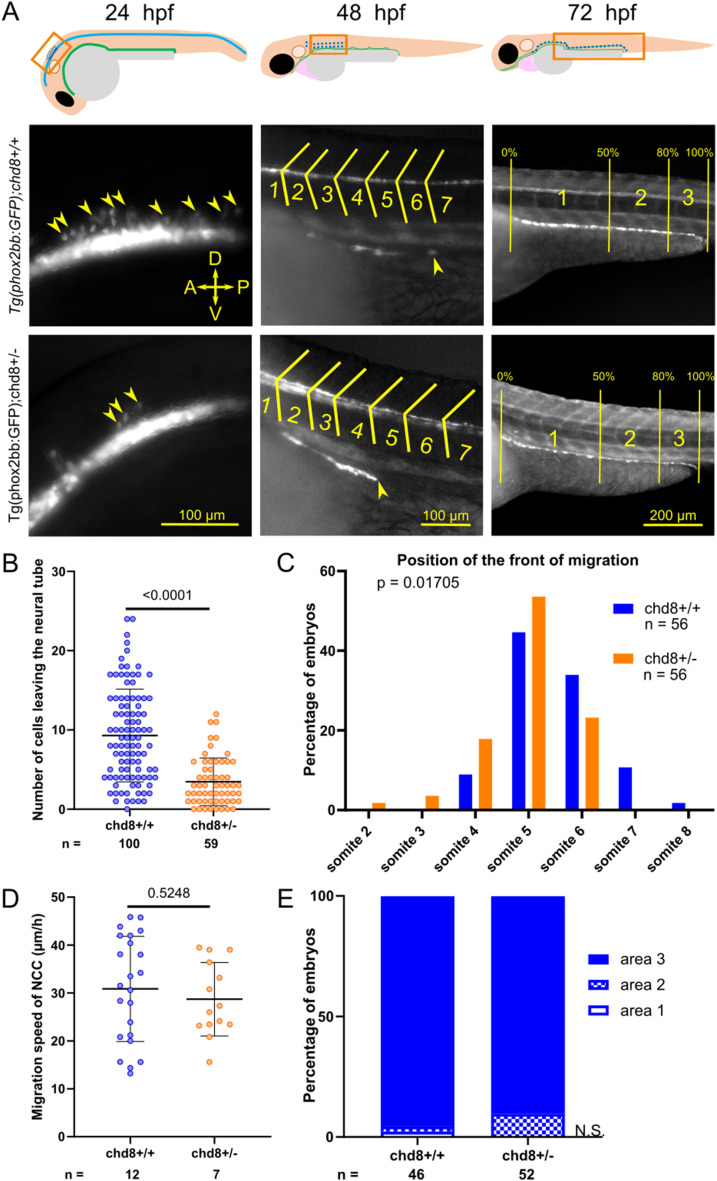
Figure 1. Heterozygous loss of chd8 leads to induction and early migration defects of vagal NCCs.
(A) Representative lateral images of Tg2(phox2bb:EGFP);chd8+/+ and Tg2(phox2bb:EGFP);chd8sa19827/+ zebrafish larvae at 24, 48, and 72 hours post-fertilization (hpf) and schematics showing the imaged areas. At 48 hpf, the somite chevrons are outlined in yellow and the front of migration is indicated by a yellow arrowhead. At 72 hpf, vertical yellow lines delimit the borders of the three following areas: area 1 represents 0–50% migration, area 2 represents 51–80% migration, and area 3 represents 81–100% migration. (B) Dot plot of the number of phox2bb-positive cells leaving the neural tube at 24 hpf. A Mann–Whitney test was conducted between pairs of conditions. (C) Histogram of the position of the front of migration of enteric NCCs at 48 hpf, using somites as morphological landmarks. Fisher’s exact test was conducted between pairs of conditions. (D) Dot plot of the measured speed of enteric NCCs between 50 and 54 hpf for each condition tested. A t test was conducted between pairs of conditions. (E) Bar graph representing qualitative scoring of the position of the front of migration of enteric NCCs at 72 hpf. Fisher’s exact test was conducted between pairs of conditions. A, anterior; P, posterior; D, dorsal; V, ventral; n, number of embryos or larvae; and n.s., non-significant.