
|
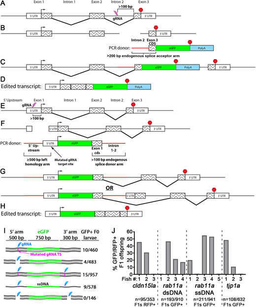
Fig. 1 Knock-in tagging in zebrafish using splice donor and acceptor arms. (A-D) C-terminal endogenous tagging strategy. An intron 5′ to the last exon is targeted for a dsDNA break (A) and integration of a PCR repair donor containing a 5′ splice acceptor element (B). mRNA splicing mediates expression of the tagged protein (C,D). (E-H) N-terminal endogenous tagging strategy. A non-coding region 5′ to the start codon is targeted for a dsDNA break (E) and integration of a PCR repair donor containing a 5′ homology arm and 3′ splice donor element (F). mRNA splicing mediates expression of the tagged protein (G,H). (I) One-cell-stage embryos were injected with different rab11a knock-in (KI) cocktails and visually screened. Cyan bolt, gRNA target site; magenta line, mutated PAM site in repair donor; ssDNA, single-stranded DNA. Numbers to the right indicate the proportion of GFP+ surviving larvae. (J) One-cell-stage embryos were injected with KI cocktails for different genes and then visually screened. GFP/RFP+ F0 fish were outcrossed, and F1 progeny were visually screened. Data plotted indicate the levels of mosaicism present in the F0 generation.