
Image
|
Figure Caption
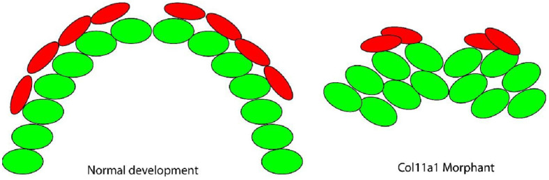
Figure 6
Model of Meckel’s cartilage mineralization in the col11a1a morphant. The organized chondrocytes (green) serve as a template for bone forming cells (red) to generate a calcified matrix. These cells extend along the template and form two bilateral mineralized rods. Therefore, perichondral cells cluster and produce abnormal mineralization at the cartilage template.
Acknowledgments
This image is the copyrighted work of the attributed author or publisher, and
ZFIN has permission only to display this image to its users.
Additional permissions should be obtained from the applicable author or publisher of the image.
Full text @ J Dev Biol