
Image
|
Figure Caption
Figure 4
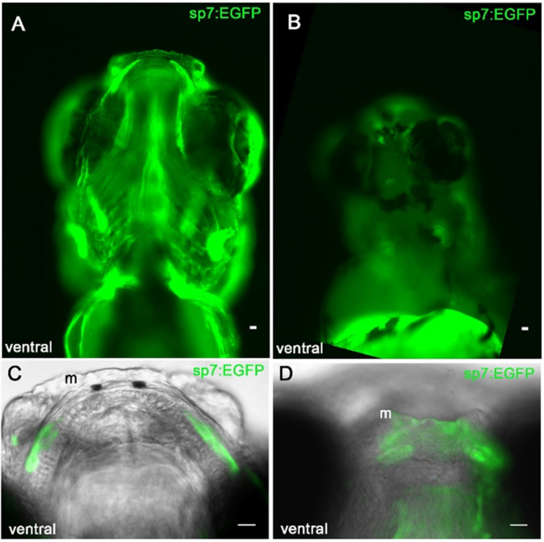
Col11a1a knockdown disrupts the organization of bone forming cells that line the Meckel’s cartilage. Ventral view of sp7:EGFP positive cells in 5 dpf control zebrafish (A,C) and morphant (B,D). sp7:EGFP expressing cells line the cartilage template in the control zebrafish (C). sp7:EGFP expressing cells form small condensations at the mediolateral cartilage but fail to occupy the location of future mineralization immediately adjacent to the Meckel’s cartilage as observed in controls (D); Scale bar 20 μm.
Figure Data
Acknowledgments
This image is the copyrighted work of the attributed author or publisher, and
ZFIN has permission only to display this image to its users.
Additional permissions should be obtained from the applicable author or publisher of the image.
Full text @ J Dev Biol