
|
Fig. 2
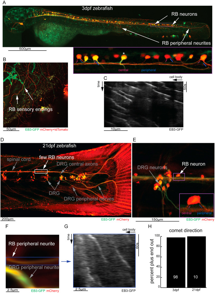
Fig. 2. Zebrafish RB neuron microtubules remain plus-end-out for the life of the cell. A. Overview of 3dpf zebrafish embryo bearing transgenic insertions of P2rx3a > EB3-GFP, P2rx3a > mCherry and Isl1[ss]>tdTomato showing the location and abundance of RB neurons at 3dpf. Enlarged image shows exit of neurites from the cell bodies in the spinal cord. B. Higher magnification image showing the morphology of RB neuron sensory endings. Transgenes are not expressed uniformly in all neurons so some cells are labeled red, others green and some both. C. A representative kymograph generated from RB sensory endings is shown. D. Overview of the posterior portion of a 21dpf zebrafish showing the position of DRG neuron cell bodies, the morphology of the peripheral nerves and the strong reduction in visible RB neurons relative to 3dpf. E. higher magnification region showing a rare visible RB neuron in a 21dpf zebrafish. F. image of a quantified portion (blue box) of RB sensory neurite in a 21dpf zebrafish. G. Sample kymograph of RB sensory neurite in a 21dpf zebrafish neuron expressing EB3-GFP H. Graph showing quantification of comet direction in 3dpf and 21dpf RB sensory neurites expressing EB3-GFP; number on column is the number of comets counted for that condition.
Reprinted from Developmental Biology, 478, Shorey, M., Rao, K., Stone, M.C., Mattie, F.J., Sagasti, A., Rolls, M.M., Microtubule organization of vertebrate sensory neurons in vivo, 1-12, Copyright (2021) with permission from Elsevier. Full text @ Dev. Biol.