
|
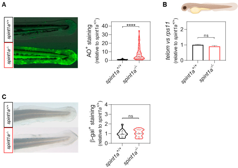
Fig. 1
Spint1a-deficient zebrafish larvae do not show premature aging at 3 dpf. (A) Representative images and quantification of the cell death levels by acridine orange (AO) staining assay in the tail of wild type (wt) and Spint1a-deficient larvae. The violin plots with the median shown as a horizontal line show the distribution of AO+ staining and are overlaid with the raw data, where each dot represents an individual. (B) The telomere length was measured in whole zebrafish larvae by qPCR using 40 ng of gDNA and determined as the telomere content relative to the single copy gene rps11. The graph shows the mean ± SEM of 25 pooled larvae (n = 25) and triplicate samples from 2 independent experiments (n = 2). (C) Quantification of the cellular senescence levels by senescence-associated b-galactosidase (SA b-gal) staining assay in the tail of wt and Spint1a-deficient larvae. The violin plots with the median shown as a horizontal line show the distribution of b-gal+ staining and are overlaid with the raw data, where each dot represents an individual. ns, non-significant; **** p < 0.0001, according to unpaired t-test with Welch’s correction (A) and unpaired t-test (B,C). Scale bars, 400 μm.