Fig. 3
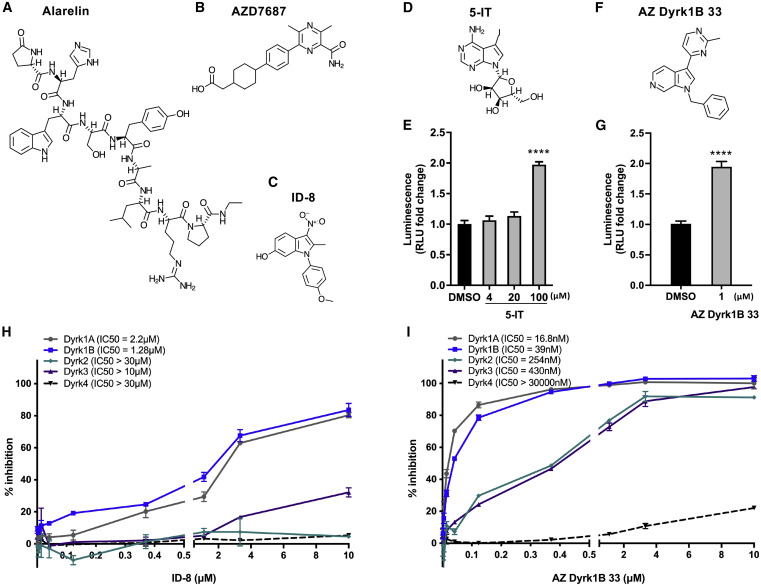
Figure 3. Structures of the hits and specificity of ID-8 and AZ Dyrk1B 33 on DYRKs (A–C) Chemical structures of the hits. Alarelin (A), AZD7687 (B), and ID-8 (C). (D–G) Chemical structures of the additional DYRK inhibitors followed up on, i.e., 5-IT (D) and AZ Dyrk1B 33 (F); and luminescence in Tg(gip:Nluc) zebrafish larvae treated from 3 to 6 days post fertilization (dpf) with 4, 20, and 100 μM 5-IT (E) or 1 μM AZ Dyrk1B 33 (G) (≥4 μM AZ Dyrk1B 33 was toxic to the zebrafish), normalized to DMSO. ∗∗∗∗p < 0.0001. (H and I) Specificity analyses of ID-8 (H) and AZ Dyrk1B 33 (I) on the DYRK-family of kinases by dose-response assessment in vitro. The SelectScreen biochemical kinase profiling assays were performed by ThermoFisher Scientific, using LanthaScreen for DYRK2 and Z′-LYTE for DYRK1A, DYRK1B, DYRK3, and DYRK4. See also Figure S2 and S3.
Image
Figure Caption
Acknowledgments
This image is the copyrighted work of the attributed author or publisher, and
ZFIN has permission only to display this image to its users.
Additional permissions should be obtained from the applicable author or publisher of the image.
Full text @ Cell Chem Biol

