Fig. 1
Image
Figure Caption
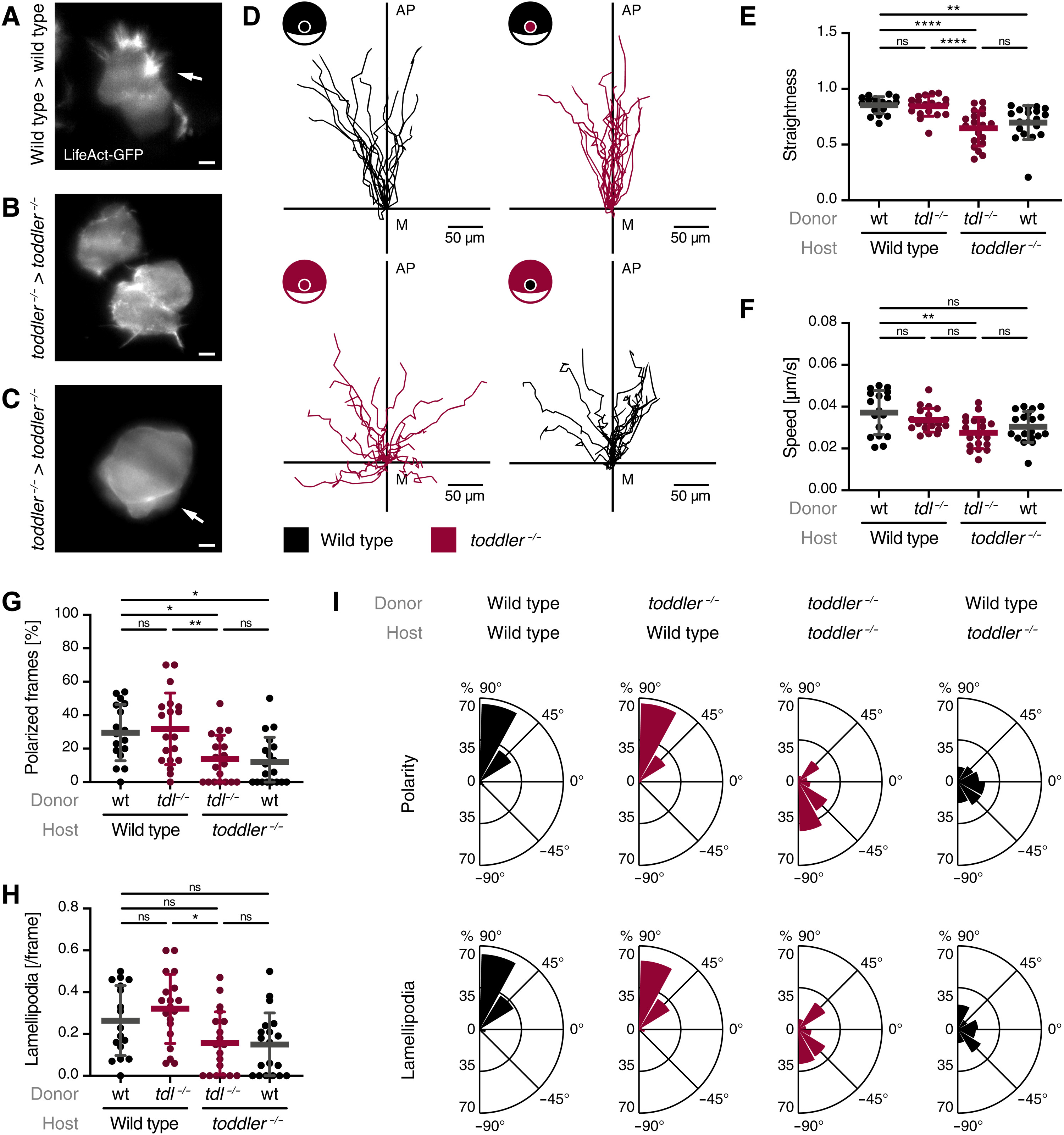
Fig. 1. Toddler acts cell non-autonomously to mediate animal pole–directed polarization and migration of mesendodermal progenitors.
LifeAct-GFP–labeled reporter cells (maximum 10 cells) were transplanted from the margin of a wild-type or toddler−/− donor embryo to the margin of a host embryo of the same or opposite genotype. (A to C) Representative light sheet microscopy images of internalized wild-type (A) or toddler−/− (B and C) reporter cells in a genotype-matched host embryo. White arrows indicate lamellipodia (A) and blebs (C). Scale bars, 10 μm. (D) Migration tracks of transplanted reporter cells [n = 19 cells, except for wild type in wild type (n = 17)]. Genotypes of donor cells and host embryos are indicated in the embryo scheme (black: wild type; red: toddler−/−). x axis = margin; y axis = animal-vegetal axis; coordinate origin = start of track. (E) Quantification of track straightness. (F) Quantification of migration speed of cells tracked in (D). (G) Quantification of cell polarity of internalized cells represented as the percentage of frames in which a cell was polarized. (H) Quantification of lamellipodia represented as average number of lamellipodia detected per frame. (I) Rose plots showing relative enrichments of orientations of polarity and lamellipodia. Data are means ± SD. Significance was determined using one-way analysis of variance (ANOVA) with multiple comparison; ****P < 0.0001; **P < 0.01; *P < 0.05; n.s., not significant. Rose plots: 90° = animal pole; 0° = ventral/dorsal; −90° = vegetal pole. All graphs are oriented with the animal pole toward the top.
Acknowledgments
This image is the copyrighted work of the attributed author or publisher, and
ZFIN has permission only to display this image to its users.
Additional permissions should be obtained from the applicable author or publisher of the image.
Full text @ Sci Adv

