
Fig. 1
|
Fig. 1
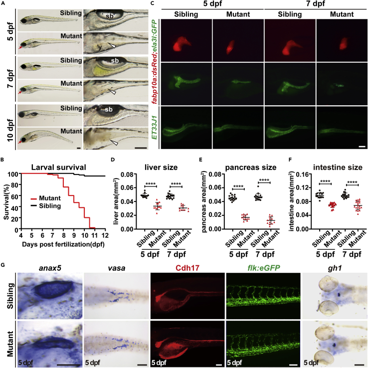
Ercc2/Xpd is crucial for zebrafish larvae development
(A) Representative images of ercc2/xpd mutants and siblings at the indicated stages. Magnified images of digestive organs are shown on the right. Red arrows indicate microphthalmia in mutants. White arrowheads point to collapse the intestine in mutants. sb, swimming bladder; y, yolk. Scale bars, 200 μm.
(B) Survival curves of ercc2/xpd mutants and siblings. n = 100 larvae each.
(C) Fluorescence images of Tg(fabp10a:dsRed; ela3l:GFP) and ET33J1 fish showing endodermal organs of ercc2/xpd mutants and siblings at 5 and 7 dpf. Lateral view, anterior to the left. Scale bar, 100 μm.
(D–F) Quantification of liver, pancreas, intestine tube area in ercc2/xpd mutants and siblings at 5 and 7 dpf. n ≥ 12. Data are presented as mean ± SD, Student’s t test, ∗∗∗∗, p < 0.0001.
(G) Whole-mount in situ hybridization and fluorescence images showing mesoderm- and ectoderm-derived organs of ercc2/xpd mutants and siblings at 5 dpf. Scale bars, 100 μm. See also Figures S1 and S2.