IMAGE

Fig 6

ID
ZDB-IMAGE-220904-27
Source
Figures for Wang et al., 2022
Image
Figure Caption

Fig 6 Reduced SCC allows for elevated mitotic recombination derived LOH.

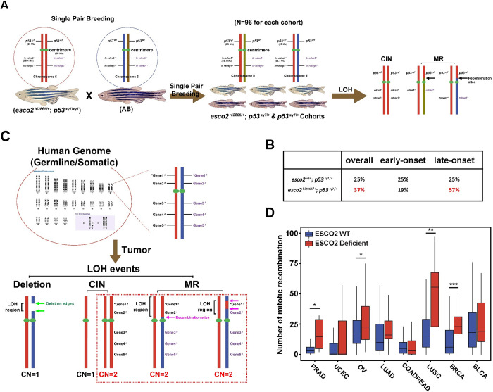
(A) Schematic of determining LOH mechanisms/types in p53 heterozygous tumors. The haplotype of nearby p53 marker in tumor and normal paired samples, cooccurring with p53 wild-type or mutant gene are labeled (left). (B) A table showing the percentage of mitotic recombination (MR) in esco2+/+; p53zy7/+ and esco2hi2865/+; p53zy7/+ tumors. (C) Schematic of determining the mitotic recombination with TCGA database. Figure A and C were created with BioRender.com. (D) The number of mitotic recombination in TCGA cancer samples with or w/o ESCO2 mutation/deletion. Genome-wide data was downloaded from NCI Genomic Data Commons. The number of WT and deficient samples in each tumor were indicated: PRAD: 481 WT + 6 Deficient; UCEC: 479 WT + 43 Deficient; OV: 383 WT + 46 Deficient; LUAD: 495 WT + 10 Deficient; COADREAD: 482 WT + 38 Deficient; LUSC: 478 WT + 8 Deficient; BRCA: 935 WT + 29 Deficient and BLCA: 393 WT + 13 Deficient. *, ** and *** indicate p<0.05, p<0.01 and p<0.001 from Mann-Whitney test.

Acknowledgments
This image is the copyrighted work of the attributed author or publisher, and ZFIN has permission only to display this image to its users. Additional permissions should be obtained from the applicable author or publisher of the image. Full text @ PLoS Genet.