
Image
|
Figure Caption
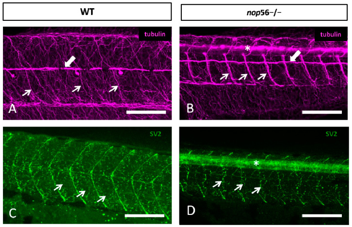
Fig. 6
(A,B) Lateral views of WT (A) and nop56−/− (B) 3.5 dpf larvae showing malformations in the neuromuscular junctions and the innervation of the trunk myomeres. All images are projections from confocal z-stacks. (A,B) Alpha-tubulin staining (magenta) showing alterations in the distribution of fibers and bundles in the trunk (thin arrows). White thick arrows in (A,B) point to posterior lateral line nerve. (C,D) SV2 (green) staining showing alterations in neuromuscular junction (arrows) and myosepta in nop56−/− compared to wild type. Asterisk in (B,D) marks the spinal cord. Scale bar: 150 μm.
Figure Data
Acknowledgments
This image is the copyrighted work of the attributed author or publisher, and
ZFIN has permission only to display this image to its users.
Additional permissions should be obtained from the applicable author or publisher of the image.
Full text @ Biomedicines