
|
Fig. 3
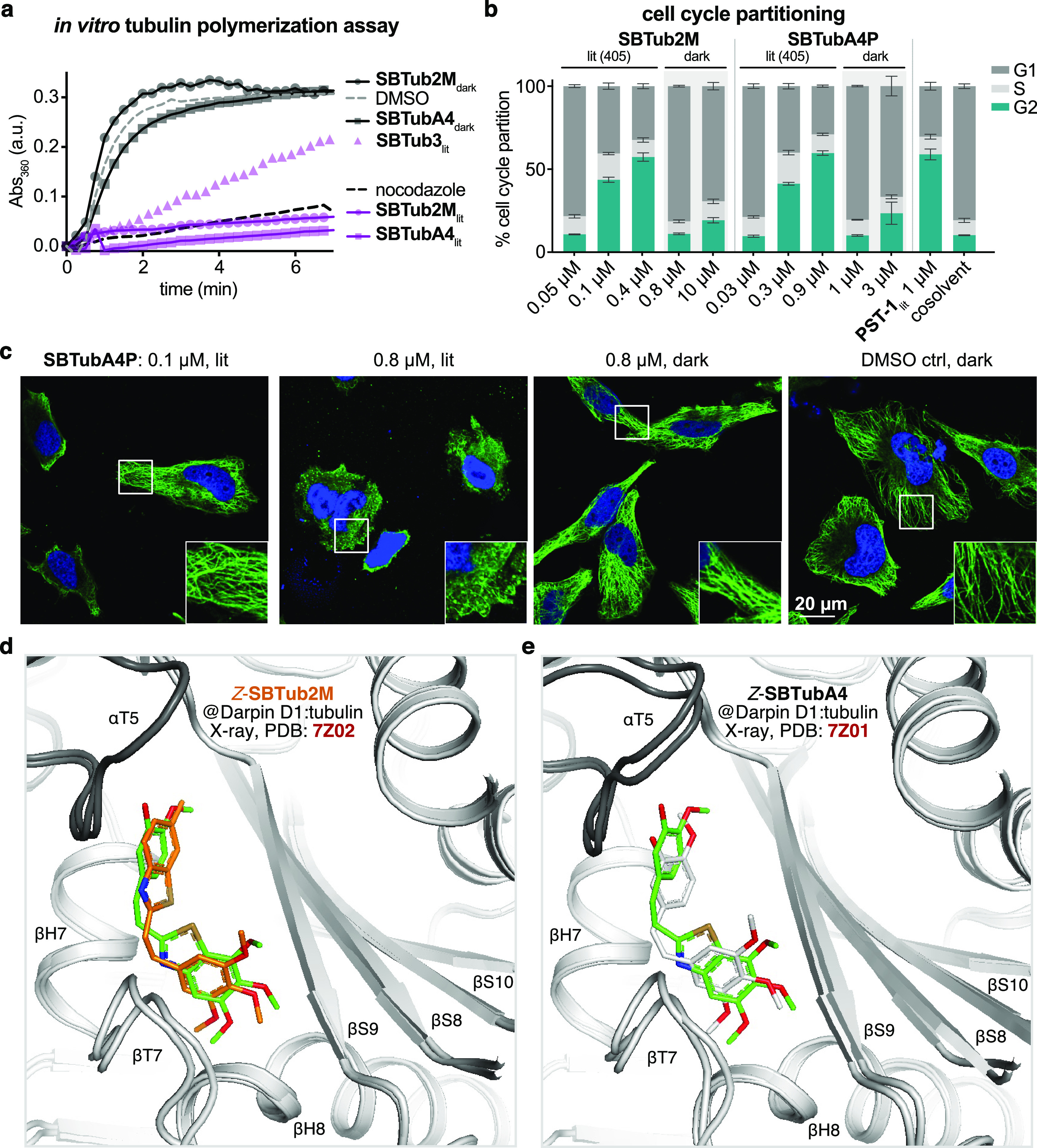
Tubulin-specific cellular mechanism. (a) SBTubs light-dependently inhibit tubulin polymerization (turbidimetric cell-free assay; absorbance mirrors the extent of polymerization; lit indicates majority-Z-SBTub (20 μM) preisomerized to PSS at 405 nm; nocodazole control at 10 μM). (b) Cell cycle analysis of Jurkat cells treated with SBTub2M/SBTubA4P matches photoswitchable reference PST-1: with significant G2/M arrest under 405 nm pulsing (lit), but without cell cycle effects in the dark (matching cosolvent controls). (c) Immunofluorescence imaging of cells treated with SBTubA4P under pulsed 405 nm illuminations (lit, mostly-Z) and in the dark (all-E), compared to cosolvent control (HeLa cells, 20 h incubation; α-tubulin in green, DNA (stained with 4′,6-diamidino-2-phenylindole, DAPI) in blue). (d) Close-up views at the colchicine-binding site of X-ray co-crystal structures (PDB 7Z01, 7Z02) of Z-SBTubA4 (green) and E-SBTub2M (orange) bound to the Darpin D1:tubulin complex (dark gray α-tubulin, light gray β-tubulin in cartoon representation; Z-SBTubA4 and Z-SBTub2M in stick representation, oxygens red, nitrogen blue, and sulfur yellow). (e) Superimposition of tubulin:CA4 (white carbons; PDB 5LYJ) and TD1:Z-SBTubA4 (PDB 7Z01) shows that lead SBTubs share the same binding site as the parent natural product CA4 (see also Figure S11).