
Image
|
Figure Caption
Fig. 3
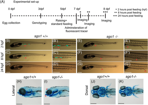
Gastrointestinal defects are not observed in sgo1 homozygous mutant larvae. (A) A schematic presentation of the intestinal transit assay experimental set up. (B–G) Representative images of 7-8 dpf larvae at different time points after feeding with fluorescently labelled tracer. Images of the same larvae at different time points are shown. Red arrowheads point to the fluorescently labelled tracer (sgo1+/+ n = 10, sgo1−/− n = 10). (H–K) Lateral and dorsal view of Alcian Blue staining in 5 dpf larvae (sgo1+/+ n = 20, sgo1−/− n = 21). Scale bars: 200 μm, taken at a magnification of 3.2× for (B–G) and at a magnification of 0.5× for (H–K). dpf: days post fertilization, hpf: hours post feeding
Figure Data
Acknowledgments
This image is the copyrighted work of the attributed author or publisher, and
ZFIN has permission only to display this image to its users.
Additional permissions should be obtained from the applicable author or publisher of the image.
Full text @ Dev. Dyn.