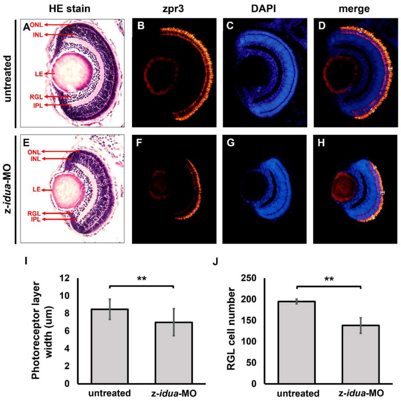
IMAGE

Figure 5

ID
ZDB-IMAGE-220801-62
Source
Figures for Lin et al., 2022
Image
Figure Caption

Figure 5

Knockdown of z-idua caused defective eyes in zebrafish embryos. Using hematoxylin and eosin staining (HE stain) and immunofluorescent staining to examine the ocular morphology of zebrafish embryos at 120 hpf. (AD) untreated control groups; (EH) z-idua-knockdown embryos. (A,E) HE staining; (B,F) immunofluorescent stain using zpr3 antibody to detect rod cells labeled in red fluorescence; (C,G) DAPI-marked nucleus labeled in blue fluorescence; (D,H) two signals merged. (I) To quantify the width of photoreceptor layer. (J) To quantify the cell number in retinal ganglion layer (RGL). All data were averaged from five independent experiments and represented as mean ± S.D. Student’s t-test was used to determine significant differences between each group (**, p < 0.01). ONL, outer nuclear layer; INL, inner nuclear layer; LE, lens; RGL, retinal ganglion layer; IPL, inner plexiform layer.

Acknowledgments
This image is the copyrighted work of the attributed author or publisher, and ZFIN has permission only to display this image to its users. Additional permissions should be obtained from the applicable author or publisher of the image. Full text @ J Pers Med